Kuopio University Hospital (KUH) in Finland is using tangential VMAT to treat breast-cancer patients and, by extension, decrease the rate of disease recurrence and increase overall survival

The radiation oncology department at Kuopio University Hospital (KUH) in eastern Finland has, for more than a decade, been treating the overwhelming majority (>98%) of its cancer patients, across diverse disease indications, using a proven combination of volumetric modulated-arc therapy (VMAT) plus daily low-dose cone-beam CT for image guidance. Zoom in a little further and it’s evident that an innovative variation on the VMAT theme – known as tangential VMAT (tVMAT) – is similarly established as the go-to treatment modality for adjuvant breast radiotherapy at KUH.
That reliance on tVMAT, which employs beam angles tangential (rather than perpendicular) to the curvature of the chest wall, is rooted in clinical upsides along multiple coordinates. Those advantages include highly conformal dose distributions for enhanced coverage of the target volume; reduced collateral damage to normal healthy tissues and adjacent organs at risk (OARs); as well as improved treatment delivery efficiency – think streamlined treatment times and lower integral dose to the rest of the body – compared with fixed-gantry intensity-modulated radiotherapy (IMRT).
Enabling technologies, clinical efficacy
If that’s the headline, what of the back-story? The pivot to a tVMAT workflow for breast radiotherapy began in 2013, when the KUH radiation oncology team took delivery of three Elekta Infinity linacs, simultaneously installing Elekta’s Monaco treatment planning system (six workstations). The KUH treatment suite also includes an Accuray CyberKnife machine (for stereotactic radiosurgery and stereotactic body radiotherapy) and a Flexitron brachytherapy unit (used mainly for gynaecological cancers).
With an addressable regional population of 250,000, the KUH radiotherapy programme sees around 1500 new patients each year, with adjuvant radiotherapy for breast cancer comprising around one-fifth of the departmental caseload. Prior to the roll-out of the Elekta linac portfolio, KUH performed breast irradiation using a 3D conformal radiotherapy (3D CRT) field-in-field technique (with planar MV imaging for image guidance integrated on the treatment machine).
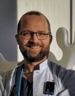
The use of 3D CRT, however, is not without its problems when it comes to whole-breast irradiation (WBI). “With the field-in-field technique, there were planning limitations for WBI related to hot and cold spots in the planning target volume [PTV],” explains Jan Seppälä, chief physicist at KUH, where he heads up a team of six medical physicists. “In some cases,” he adds, “target coverage was also compromised due to heart or lung dose constraints.”
Fast-forward and it’s clear that the wholesale shift to tVMAT with daily cone-beam CT imaging has been a game-changer for adjuvant breast radiotherapy at KUH. Although the clinical and workflow benefits of conventional VMAT techniques are also accrued across prostate, head-and-neck, lung and other common disease indications, Seppälä and colleagues have made breast-cancer treatment a long-term area of study when building the evidence base for VMAT’s clinical efficacy.
“We have found that, with proper optimization constraints and beam set-up in the Monaco treatment planning system, tVMAT can reduce doses to the heart, coronary arteries and ipsilateral lung,” Seppälä explains. “The technique also enhances dose distributions greatly – reducing hotspots, improving target-volume dose coverage, while avoiding high-dose irradiation of healthy tissue as well as a low dose bath.” All of which translates into fewer reported side-effects, including breast fibrosis, changes in breast appearance, and late pulmonary and cardiovascular complications.

Operationally, the total treatment time for breast tVMAT – including patient set-up, cone-beam CT imaging, image matching and treatment delivery – is approximately 10 minutes without breath-hold and about 15 minutes with breath-hold. The average beam-on time is less than two minutes.
“We use daily cone-beam CT image guidance for every patient, with the imaging dose optimized to be as low as possible in each case,” notes Seppälä. The cone-beam CT highlights any breast deformations or anatomical changes during the treatment course, allowing the team to replan if there are large [>1 cm] systematic changes on the patient surface likely to affect the dose distributions.
It’s all about outcomes
Meanwhile, it’s clear that toxicity and cosmetic outcomes following breast radiotherapy have improved greatly at KUH over the past decade – evidenced in a small-scale study by Seppälä’s team and colleagues at the University of Eastern Finland. Their data, featured in a poster presentation at last year’s ESTRO Annual Meeting, provide a comparative toxicity analysis of 239 left- or right-sided breast-cancer patients, with one cohort treated with tVMAT (in 2018) and the other cohort treated with 3D CRT (in 2011).
In summary, the patients treated in 2018 with the tVMAT technique exhibited less acute toxicities – redness of skin, dermatitis and symptoms of hypoesthesia (numbness) – versus the patients treated in 2011 with 3D CRT. Late overall toxicity was also lower, and the late cosmetic results better, in the 2018 patient group. “With tVMAT,” says Seppälä, “we have much less skin toxicity than we used to have with previous 3D CRT techniques. What we are still lacking, however, is the systematic and granular capture of patient-reported outcomes or daily images of the patient’s skin after each fraction.”
For Seppälä, comprehensive analysis of those patient-reported quality-of-life metrics is the “missing piece of the jigsaw” – and, ultimately, fundamental to continuous improvement of the tVMAT treatment programme at KUH. A case in point is the ongoing shift to ultra-hypofractionation treatment schemes in breast radiotherapy, with some KUH patients now receiving as few as five fractions (x5.2 Gy) as opposed to 15 (x2.67 Gy) fractions per the norm to date.
To support this effort, work is under way to evaluate the clinical implementation of Elekta ONE Patient Companion, powered by Kaiku Health, a system providing patient-reported outcomes monitoring and intelligent symptom-tracking for cancer clinics. “This software tool would enable us to capture real-world outcome data directly from patients,” says Seppälä. “Those data are key for quantifying success, such as the correlation of cosmetic outcomes with a change in fractionation scheme.”
Meanwhile, machine-learning innovation is another priority on KUH’s tVMAT development roadmap, with the medical physics team in the process of implementing AI-based dose predictions to inform treatment planning on an individualized patient basis. The driver here is the push for more unified, standardized dose distributions as well as workflow efficiencies to streamline patient throughput.
“We are doing some treatment planning automation – mainly on the optimization side,” Seppälä concludes. “The challenge is to push the optimization system to its limit to ensure low doses to critical structures like the heart and ipsilateral lung. By doing so, we can deliver at-scale enhancements to the overall quality and consistency of our treatment planning in Monaco.”
Read more
Elekta Unity: CMM innovation opens the way to real-time tracking, online plan adaptation


