Cherenkov imaging enables real-time visualization of radiation beams on a patient’s body and provides a means to evaluate the accuracy of radiotherapy delivery. Researchers in China have now developed a way to improve the quality of Cherenkov images using a flexible, non-toxic sheet of carbon quantum dots (cQDs) attached to the patient.
Cherenkov light is produced when charged particles travel at a speed greater than the phase velocity of light in tissue. The signal intensity is proportional to the delivered radiation dose, revealing the precise dose that’s delivered during treatment. The optical imaging technique offers high spatial resolution, high sensitivity and fast imaging speed compared with conventional methods of radiation dose measurement.
The intensity of Cherenkov emission is low, however, and the emitted photons are scattered and absorbed by tissue. Because of this, standard charge-coupled device (CCD) cameras have difficulty collecting the signal. Instead, more expensive intensified CMOS/CCD cameras are being used.

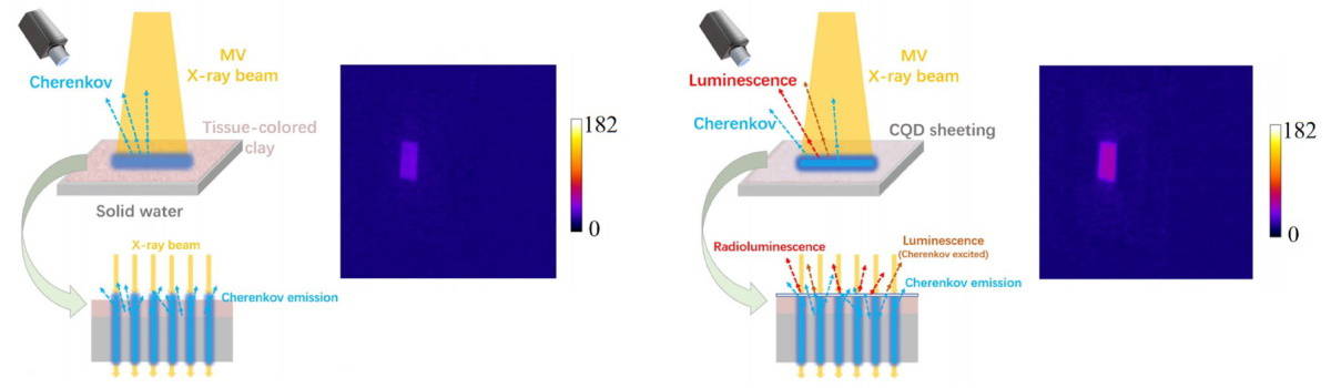
The cQDs have absorption spectra that overlap with the Cherenkov emission spectra; they then emit luminescence at longer wavelengths. The cQD sheeting, developed and tested at the Department of Nuclear Science and Technology of Nanjing University of Aeronautics and Astronautics, can therefore be used to shift the Cherenkov emission to match the optimal wavelength of a CCD camera’s sensitive detection region.
With the cQD sheeting in place, the optical emission is composed of Cherenkov photons generated in the superficial surface of the tissue, fluorescence excited by the Cherenkov photons, and the radioluminescence generated in the cQDs. This increases the total optical signal and improves the image quality and signal-to-noise ratio (SNR) of the acquired images.
Principal investigator Changran Geng and colleagues created the cQD sheeting using a solution of 10 nm-diameter cQDs and UV-curable adhesive. This mixture was spin-coated onto a substrate coated with plastic sheeting and solidified with a UV lamp. The plastic substrate ensures that the scintillation material does not directly contact the skin.
The resulting cQD sheeting had a thickness of 222±5 µm and a diameter of 15 cm, and was flexible enough to conform to the patient’s surface. The team note that the cQD sheeting is almost transparent and does not block the Cherenkov emission from tissues.
Reporting their findings in Medical Physics, the researchers initially tested the cQD sheeting on a solid water slab covered with a 2 mm layer of light coloured skin-toned clay to mimic the optical properties of skin. They evaluated the relationship between optical intensity and delivered dose using cQD concentrations of 0, 0.05 and 0.1 mg/ml, delivered doses of 100–500 MU, and 6 and 10 MV beams. They observed a linear relationship between optical intensity and dose for both 6 and 10 MV photons. Adding the cQD sheeting more than doubled the SNR in both cases.
The team then examined the performance of the cQD sheeting on an anthropomorphic phantom using different radiotherapy materials and various ambient light sources. Light emission from the surface of the various materials was over 60% higher with cQD sheeting than without. Specifically, the average optical intensity increased by about 69.25%, 63.72% and 61.78% when adding cQD sheeting to bolus, mask sample, and a combination of bolus and mask, respectively. The corresponding SNRs improved by about 62.78%, 56.77% and 68.80%.
Under ambient light from a red LED, optical images with a SNR of greater than 5 could be obtained through the sheeting. Adding a band-pass filter increased the SNR by about 98.85%.
“Through a combination of cQD sheeting and corresponding filter, the light intensity and SNR of optical images can be increased significantly,” the researchers write. “This sheds new light on the promotion of the clinical application of optical imaging to visualize the beam in radiotherapy with a more rapid and less expensive image acquisition process.”

Cherenkov imaging for visualizing radiotherapy: one year of clinical use
Geng tells Physics World that the team is actively continuing its research in many ways. One example is investigating Cherenkov imaging for use with electron beam radiotherapy of keloids, benign fibrous lesions arising from an abnormal healing response.
“Some studies have indicated that post-operative electron beam radiotherapy can reduce the rates of keloid recurrence,” Geng explains. “However, inaccurate deliveries are commonly associated with the variation of electron beam parameters, as well as patient’s setup uncertainties or respiratory movements. These can lead to insufficient or excessive dose at the mismatched adjacent fields, potentially causing tissue damage to normal skin or keloid recurrence. We are trying to use Cherenkov imaging technology with cQD sheeting to measure matching of adjacent radiation fields delivered during keloid electron radiotherapy in real-time.”