A monochromatic X-ray source under development for breast cancer screening can produce high-quality phantom images at significantly lower radiation dose and with greater sensitivity than conventional mammography systems. This low-dose technology could represent a paradigm change for mammography, making these exams safer for women by exposing them to less radiation dose and better identifying suspicious abnormalities, especially in dense breasts.
Developers at Imagine Scientific report that their prototype system reduces radiation dose by a factor of five to 10 times for the same signal-to-noise ratio (SNR) as a conventional mammography system. They tested the system on breast phantoms of varying thickness containing a variety of test objects. The system is being designed for both conventional 2D digital mammography and 3D digital breast tomosynthesis (DBT).
Conventional radiography systems use multi-wavelength X-ray emission, which extends over a broad energy band. This creates a less optimal image than an extremely narrow bandwidth beam does, and exposes patients to a higher level of radiation to obtain a diagnostic-quality image. The advantages of monochromatic X-ray radiation to improve clinical imaging are well known, but its generation requires crystal or multilayer monochromators coupled to either traditional broadband X-ray tubes or large synchrotron facilities. Both are impractical for routine clinical use.

Company founder Eric Silver and colleagues have developed a prototype X-ray generator that sequentially combines two X-ray emission processes to generate monochromatic X-ray beams suitable for medical imaging. The two-stage process includes bombarding a metallic target with high-energy electrons to emit broadband bremsstrahlung X-rays, which subsequently irradiate a second target that emits monochromatic X-rays via fluorescence. Additional proprietary geometric techniques produce a beam of monochromatic X-rays with sufficient intensity and large enough field-of-view for routine clinical imaging.
The prototype X-ray tube, which is comparable in size to a conventional X-ray tube, uses the inside surface of a conically-shaped annular ring to concentrate the broadband X-ray energies onto a compact, thin-foil, secondary metallic target placed at the centre of the ring. This secondary target subsequently emits monochromatic X-rays via fluorescence, with an energy determined by the elemental composition of the compact target. The team describe the design in detail in Medical Physics.
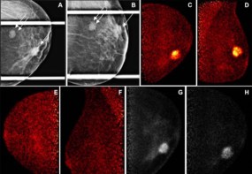
For their validation study, the researchers recorded images of four breast phantoms with thicknesses of 4.1, 4.5, 7.1 and 9.0 cm, using the prototype and a conventional 2D mammography system. The phantoms contained high- and low-contrast masses and microcalcifications, as well as regions of differing breast densities. The team compared image quality as a function of radiation dose using the measured SNR values.
The researchers reported that tissue fibres, microcalcifications and low-contrast lesions were observable in both sets of phantom images. However, the conventional system required 4.5 times the dose for the 1.0 and 0.75 mm thick lesions, and six to eight times the dose for 0.5 to 0.2 mm thick lesions to produce the same SNR as that of the monochromatic system.
“The high SNRs for very thick breast phantoms provide evidence that screening with less breast compression is possible while maintaining high image quality, helping to improve patient comfort,” write the authors. They also report that “contrast-enhanced digital mammography with monochromatic X-rays was shown to provide a simpler and more effective technique at substantially lower radiation dose”.
The technology may potentially reduce unnecessary negative biopsies and avoid the need for expensive follow-up imaging, such as breast MRI and molecular breast imaging.
“Our objective is to replace conventional broadband X-ray tubes used in 2D mammography and 3D DBT with our monochromatic X-ray source,” says Silver. “The same improvements in SNR, detectability, image quality and lower radiation dose that we demonstrated for our 2D measurements apply to 3D DBT as well.”
“The technology we are developing will provide unambiguous results, 85% lower radiation dose, examinations without painful breast compression, and the ability to perform chemical analysis of tissue in vivo.” he adds. “Any medical application that uses a conventional X-ray tube will benefit from using monochromatic radiation. The additional disciplines we are addressing include paediatric, bone, lung, heart, thyroid, prostate and dental imaging.”
Silver tells Physics World that a second-generation monochromatic X-ray tube with at least ten times higher power is currently in development to reduce exposure time, and that automatic replacement of the secondary fluorescence target is a priority. Image Scientific is planning to sponsor a series of clinical studies in leading medical centres. These include a pilot study comparing conventional and monochromatic mammography of women with dense breasts, to be followed by a large, multi-institution clinical study in 2022.