Physics World, which was founded in 1988, turns 30 in October and as part of our anniversary celebrations we’ll be publishing a special issue of our magazine. In that issue, we’ll be looking back at the key topics in physics 30 years ago, seeing where those fields are now and then contemplating where those endeavours will take us over the next 30 years.
In preparation for the special issue, I travelled to Imperial College London today to interview Julia Higgins, president of the Institute of Physics, which publishes Physics World. Higgins, who originally studied physics at the University of Oxford, went on to have a glittering career in polymer science, spending almost two decades as a professor in the chemical-engineering department at Imperial.
She has also served in a huge number of roles, including president of the British Association for the Advancement of Science, co-founder of the Athena Project to encourage women into science and chair of the Engineering and Physical Sciences Research Council. She was even a trustee for the National Gallery
Sitting in her sweltering office on fifth floor of the department, I quizzed Higgins on everything from the highlights of her career over the last 30 years to her views on research funding, education, Brexit, diversity and more. You’ll have to wait for the October issue to read her views in full, but I don’t think she’ll mind me revealing here the role she enjoyed the most.
It was her stint as “foreign secretary” of the Royal Society, which saw her travel on numerous trips around the world. Now it’s no secret that foreign trips are one of the many perks of being a scientist, but as I left Imperial I spotted two mentions of another physicist from the college who’s also been on her travels.
That person is Melanie Windridge, who completed a PhD in plasma physics at Imperial in 2009 and is now an author, broadcaster, and fusion consultant for Tokamak Energy. Windridge, who in 2014 wrote a book Aurora based on a trip in search of the Northern Lights, earlier this year climbed Mount Everest. Her trip was covered in a recent issue of the Imperial magazine Reporter – copies of which littered the campus – and was also mentioned on a big screen in the entrance foyer to the college.
In fact, Windridge, who is a visiting fellow in Imperial’s physics department, has also written a full-length feature about her adventure for Physics World. Set to appear in the September issue of the magazine, she describes how science and technology are vital to anyone climbing Everest. She’ll also be writing for the 30th-anniversary issue, this time on the prospects for fusion energy.
Now I don’t know if this analogy has been made before, but I reckon there are lots of parallels between doing research and climbing a mountain. Both are big challenges. Both need careful planning. Both rely on team-work, communication and co-operation. Both require a good understanding of existing techniques and knowledge. And both are massively risky endeavours in which the end-goal is often visible but failure is ever-present.
But as both Higgins and Windridge I am sure will attest, the rewards of mountaineering and scientific research can be immense.
Computer simulations done by physicists in Switzerland and the US suggest that “invisible anchors” can trap particles as they move with a fluid through T-junctions. The regions in which particles are trapped can comprise large parts of a junction and the research could lead to the design of microfluidic systems that are designed specifically to trap and process particles of interest.
T-junctions and other connections between fluid-carrying pipes are ubiquitous in the built-up world – and can play important roles in industrial processes. While it seems reasonable to think that liquid flow will pull small particles through a junction, in reality particles are known to become trapped in large regions of a junction. This can lead to a wide range of problems such as clogged pipes. On the positive side, this trapping effect is being used to create microfluidic systems for processing particles.
Since 2014, Howard Stone of Princeton University and colleagues have been studying this effect by doing experiments in which hollow glass beads are carried by fluids of different viscosities through a variety of junctions. They found that these beads – which have lower density than the fluids – can become trapped in junctions under a wide range of experimental conditions.
Vortex breakdown
The team suspected that the trapping is caused by a phenomenon called “vortex breakdown”. This is an abrupt disruption of the smooth flow of a fluid, which can occur as the fluid accelerates around the sharp bend of a junction. This creates vortices and regions of zero flow where particles can be trapped.
Stone and colleagues identified this mechanism in their earlier work, but remained puzzled because their calculations suggested that the trapping regions were much smaller than seen in experiments. Now, Stone has teamed up with David Oettinger and George Haller of ETH Zurich and Jesse Ault of Oak Ridge National Laboratory to do much more detailed calculations that consider how individual particles interact with the fluid – and how this affects their motion within a T-junction.
The team’s computer simulations revealed much larger trapping regions that resemble a ship’s anchor. In some cases, 25% of the cross-section of the input pipe led to regions in which particles would become trapped.
Gaining a better understanding of the trapping process could lead to a wide range of applications including minimizing the build-up of bubbles within liquid distribution systems and processing materials in microfluidic devices. Describing their latest results in Physical Review Letters, Stone and colleagues highlight the microfluidic production of single-layer lipid vesicles as one important potential application.
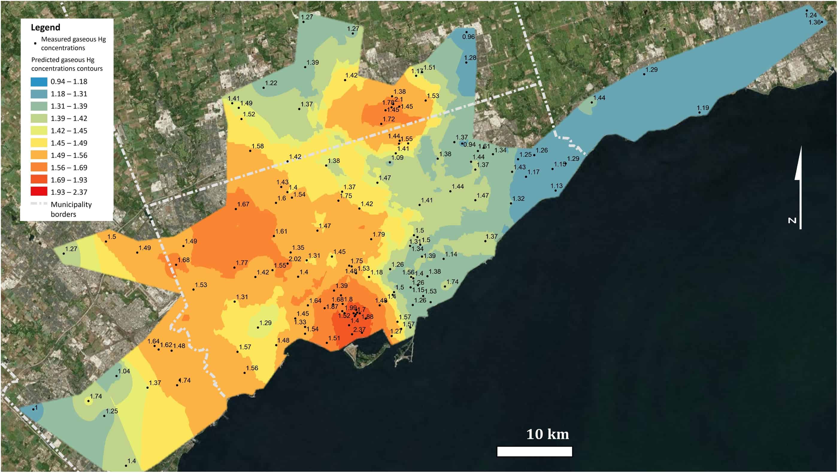
Measured and predicted gaseous mercury concentrations (ng m−3) averaged over a period of 4–6 weeks in July/August 2016 in the Greater Toronto Area. (Courtesy: Identifying and evaluating urban mercury emission sources through passive sampler-based mapping of atmospheric concentrations David S McLagan et al 2018 Environ. Res. Lett. 13 074008 doi:10.1088/1748-9326/aac8e6)
Urban areas emit mercury from fossil fuel combustion, metal manufacturing, cement and caustic soda production, medical and industrial waste, and cremations. Other sources may be significant too but have been hard to quantify. The UN’s Minamata Convention on Mercury adopted in 2013 calls for complete emissions inventories and better monitoring networks for mercury.
“Up to now do we really have a great handle on what these fugitive emissions could be in a city? Not really,” says Carl Mitchell of the University of Toronto Scarborough, Canada. “The assumption is that the vast majority of emissions to the environment are from the major known point sources.” Sources like coal-fired electricity plants and factories.
Such emissions are of serious concern because exposure to mercury causes severe neurological damage. Mercury itself is highly volatile, meaning it evaporates like water into the atmosphere. Once there it travels the globe; pollution from Europe, for example, can contaminate food chains in Canada.
To understand the role of fugitive emissions, Mitchell and his colleagues deployed 145 small passive air samplers across the Greater Toronto Area in July and August 2016. Each sampler cost about $10, didn’t require an energy source or compressed gases, and provided time-averaged concentration data.
A total of 58 staff and students of the University of Toronto Scarborough put a sampler at their home; at other sites the team attached samplers to a utility pole or tree. 43 of the samplers were near potential mercury sources – waste/recycling centres accepting mercury-containing products, crematoria, which may release mercury from dental amalgams, and hospital/dental facilities.
The team also deployed samplers in the summer of 2016 or 2017 near known sources of mercury – facilities within 100 km of downtown Toronto that report their atmospheric mercury emissions to Canada’s National Pollutant Release Inventory, including a waste facility, two steel plants, a cement producer and a wastewater treatment plant.
In this way, the team created a more detailed map of the emission sources in the Greater Toronto Area, and identified several small fugitive emitters in the city, including waste and recycling, and hospital/dental facilities.
Some results were surprising. A couple of sites reported by government sources to be high mercury emitters were, according to the samplers, low emitters. And other presumed low emitters measured higher concentrations of gaseous mercury than expected. Mitchell believes this speaks to the need for better methods and accuracy in government reporting on atmospheric mercury. The good news, according to the researchers, is that the passive samplers the team employed may be the perfect solution.
“Without this instrument, I feel, it is really difficult to otherwise get this information,” said Mitchell. Traditionally, scientists have deployed more expensive, active instruments at just a few sites in urban areas to monitor atmospheric mercury over the long term.
The samplers are cheap and easy to use while still providing accurate measurements. To begin to address fugitive emissions, explains Mitchell, “you need to be able to tell the difference between two relatively small numbers” – the source emission level and the background level – “and the really powerful thing we observed was we could statistically see those differences”.
Comparing the team’s findings with previously published studies, Mitchell says, “we’ve seen a definite reduction in the amount of mercury in the air over the past ten years or more”. He credits this to mitigation efforts already in place in Canada, such as the elimination of all coal-fired power in Toronto’s home province of Ontario.
According to Mitchell this work also provides an interesting case study for the use of passive samplers in the challenging urban environments of India and China, where they use more mercury in industry. “You could do a similar study there,” he says, “and it might actually be quite eye-opening.”
A vaccine made from double-layered polypeptide nanoparticles appears to protect mice against flu. The vaccine, delivered using dissolvable microneedle patches, might be adapted to treat other pathogens too.
Seasonal flu vaccines must be updated every year by predicting which strains will be the most common. Mismatched vaccines are obviously not very efficient and provide only limited protection, which increases the risk of a flu epidemic. Ideally, universal flu vaccines that are active against a broad range of influenza viruses would be better, but developing these is no easy task.
Researchers led by Bao-Zhong Wang of Georgia State University in the US have now built on recent studies that showed that the internal influenza protein nucleoprotein (NP) can induce T-cell immune responses in cells. Each double layer in their new nanoparticle vaccine contains a core made of peptides from this nucleoprotein. The outer coating of the nanoparticle contains four peptides from the ectodomain epitopes of the influenza A M2 protein (M2e). This protein is a promising target for “universal” flu vaccines because it exists in most human seasonal influenza A viruses.
Long-lasting immunity
Wang and colleagues administered their vaccine using a dissolvable microneedle patch and found that it provided mice that had been exposed to the influenza A virus with long-lasting immunity. The nanoparticles appear to trigger immune responses not only in T lymphocytes, as expected, but B lymphocytes as well, so providing broad cross-protection.
The team, which includes researchers from the Georgia Institute of Technology, Emory University, both in the US, and Henan Normal University in China, says that it now plans to improve its vaccine by adding the “stalk” (the inside portion of the influenza virus’ surface protein) to its coating. Seasonal flu vaccines provide immune protection by targeting the head of this surface protein (known as hemagglutinin), but this head varies greatly from one virus strain to the next, explain the researchers. The stalk, on the contrary, does not vary so much and so might be exploited to develop a universal protective vaccine, they say.
Their technique, which is detailed in PNAS 10.1073/pnas.1805713115, might also be used to develop vaccines for other pathogens, and perhaps even cancers, they add.
Walk into any modern physics laboratory and you’ll see all kinds of hi-tech instruments. There are spectrometers, microscopes, oscilloscopes and diffractometers all spitting out data, spectra and images. Apart from being expensive, the main problem with these “black-box” instruments is that they can’t be fully inspected or customized. If they break, you often have to pay engineers to come and fix them.
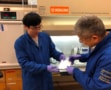
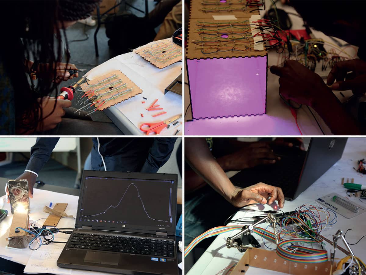
But what if you could make your own equipment? This is the principle behind the open-science hardware movement, which lets people make, modify and share hardware for scientific use. By sharing design blueprints and using 3D printers, equipment can be made quickly and cheaply. The idea has caught the imagination of many researchers, but for scientists in Africa and other parts of the developing world, open-science hardware is a lifeline that could benefit their teaching and research.
(Courtesy: Top row: TReND in Africa; Open-Labware.net. Middle row: TReND in Africa. Bottom row: TReND in Africa; Richard Bowman)
The trend for open-source hardware kicked off in the mid-2000s. It evolved from the “maker movement” in the US where DIY culture merged with hacker culture, in which members of the IT community collectively modified and developed code to improve software systems. At the core of the maker movement was the philosophy that making it easier for individuals to create things themselves could lead to a new era of micro-manufacturing and end the monopoly of mass-manufacturing. The device to enable this was the 3D printer, which was becoming cheap enough for the consumer market. People could start sharing designs thanks to online depositories such as Thingiverse, and could 3D-print them from plastic using the Lego polymer, acrylnitrile butadiene styrene (ABS), or biomass-derived polylactic acid (PLA).
The maker movement soon found its way to the sciences, but it wasn’t quite as simple as making other, non-scientific things according to Jenny Molloy, an early proponent of open science and a research fellow at the University of Cambridge, UK. “[General makers] didn’t tend to have to deal with the quality assurance, adherence to standards, calibration and reproducibility that is needed in science,” she says.
The open-source concept was not entirely new to science either. In the 1990s Linus Torvalds at the University of Helsinki, Finland, designed the Linux operating system, which provided an underlying source code that could be used, modified and distributed by anyone. Now it’s widely used in servers, software and firmware, including Android smartphones, TiVo digital video recorders and satellite navigation systems for vehicles. Its success has often been attributed to its collaborative open-source development because it provides speedy problem solving.
A joy in sharing
So could a similar approach work for scientific hardware design? One of the first organizations to test this was CERN in Switzerland. “Most printed circuit boards designed at CERN are now made as open hardware,” says Molloy. “For them it a very pragmatic approach.” Since 2009 CERN’s open hardware repository (OHR) enables developers to avoid duplication and review work. They have also created their own open-hardware licence to provide a legal framework surrounding this technology exchange.
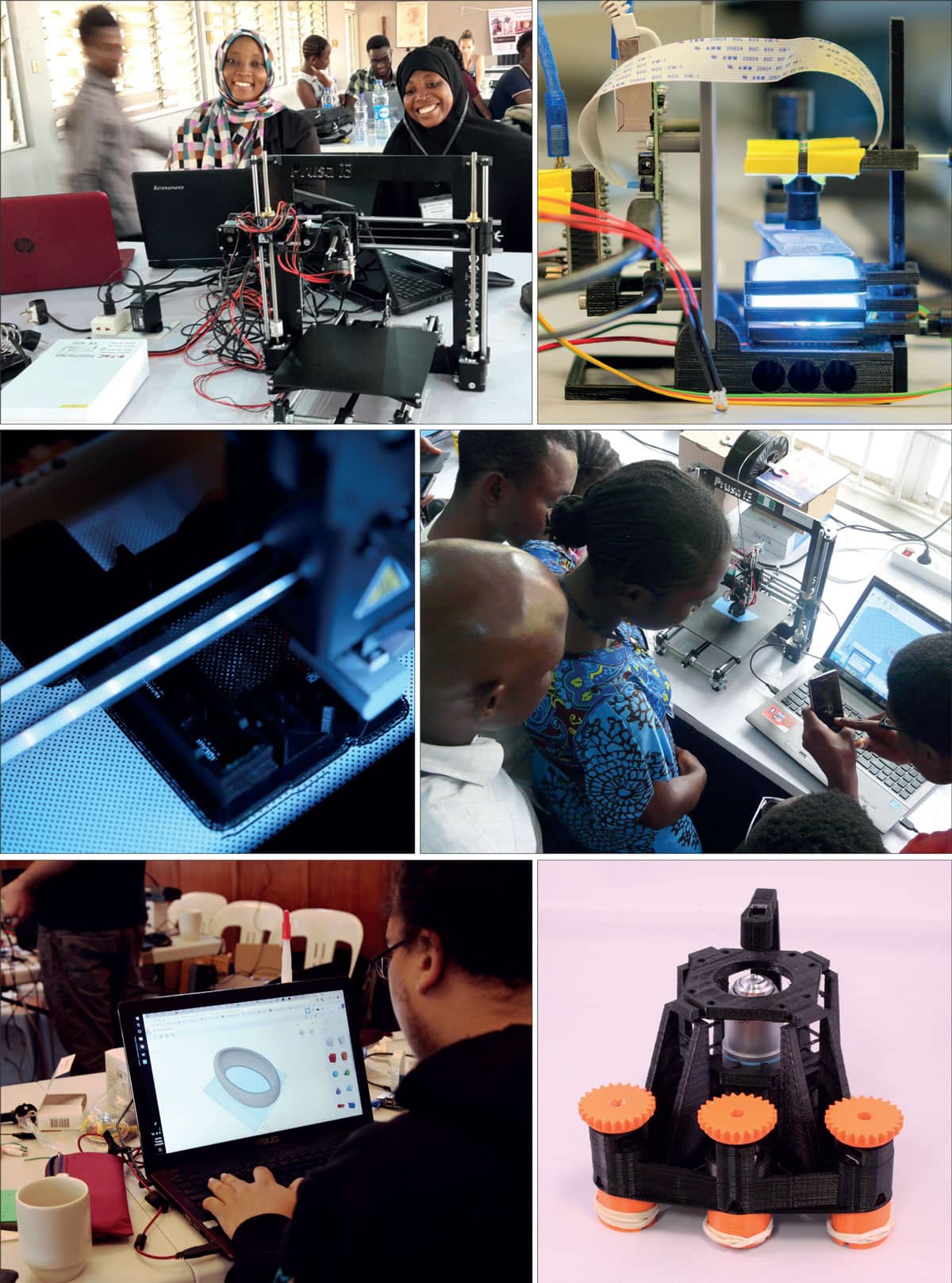
Another proponent of open-source hardware is Tom Baden, a neuroscientist at the University of Sussex in the UK, who became involved in 2012 after buying a 3D printer. He admits he did “not really have much of a plan” and started printing objects using designs downloaded from the Internet. Keen to design his own things, he took inspiration from a crude micropipette. “I thought maybe I can take the basic idea and design one that is a bit more precise,” Baden recalls.
Having done that, he then set up the website Open-Labware.net to test online reaction to his designs. It now offers numerous open-science hardware designs and modifications of existing equipment. Very popular designs, such as the pipette, can be downloaded around 30,000 times. “There is a joy in sharing,” Baden says.
Printed pipettes: Tom Baden refined a crude design for a micropipette. (Courtesy: Open-Labware.net)
As well as 3D printers, the other important development in the open-hardware community is the emergence in recent years of cheap, powerful electronics such as Arduino microcontrollers, Raspberry Pi computers and custom integrated circuits. Therefore, the designs available from online repositories can range from tiny accessories, like pipettes, to complex machines. “There are research scientists in physics who, on the side, build some really quite complicated machines, for example scanning laser microscopes,” says Baden. A popular design is a polymerase chain reaction (PCR) thermal cycler used by all molecular biology labs to amplify DNA. “It costs a few thousand dollars, but it’s basically just a heating and cooling device with a timer, so you can imagine it is fairly easy conceptually to build,” he adds.
Some devices, meanwhile, are being built primarily for educational purposes, rather than research. They include a Lego-based low-cost atomic force microscope (AFM), which was built in 2013 by an international student group as part of the Lego-sponsored LEGO2NANO event. The design now forms the basis of OpenAFM – a not-for-profit start-up to encourage other schools to build these devices.
Richard Bowman, an optics physicist at the University of Bath, UK, has likewise developed an open-source microscope called the OpenFlexure microscope that could be used in schools. The 3D-printed light microscope uses a modified web cam as the optics and includes a mechanical stage that allows sub-micron positioning of a sample and lens. Bowman’s design has been shared openly on the software development platform GitHub and can also be easily modified and customized.
“The idea of being able to share hardware in a similar way to how you share software really appealed to me,” he says. “The most basic version [of the OpenFlexure microscope], is definitely in the same ball park as a basic school microscope – it’s good for seeing single human cells.” But for Bowman there is a wider appeal. “I can flip a few switches in the design and get something that uses a proper objective lens and other optics, meaning that the images produced should be more or less equivalent to the sorts of microscopes that are being used in hospitals in the developing world.”
International community
Bowman’s desire to make something that could be used in developing nations is a common theme among members of the open-science hardware community. “Experimental science is really held back [in the developing world] by not being able to get hardware and, when they have hardware, not being able to service it and maintain it,” says Bowman. “So the hardware ends up sitting on a pedestal in the corner of the lab, not being used except to show off to visitors because actually it’s impossible for them to keep it working.” Open-science hardware could therefore help to bypass these problems.
Spreading science: TReND in Africa holds workshops to teach people how to make their own hardware. (Courtesy: All images: TReND in Africa)
In 2010 Baden founded a charity called TReND in Africa (Teaching and Research in Neuroscience for Development) with fellow neuroscientist Lucia Prieto-Godino, who is now at the Crick Institute in London. It supports neuroscience (and often other disciplines) in African universities by providing training in open-science hardware.
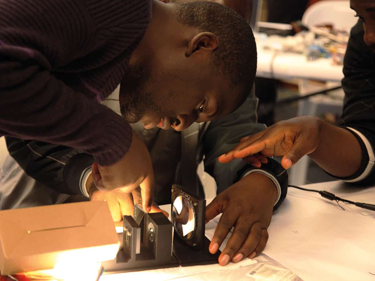
André Maia Chagas, who is a project leader for TReND in Africa, sees the task as one of developing local expertise and capabilities. “We show people that with very basic knowledge of electronics and 3D printing, they can build their own lab equipment, so we have been trying to show people that it’s something everyone can do,” says Maia Chagas. He does this by organizing local workshops, including a two-week event that was held at the University of Ibadan, Nigeria, in July 2017, and had 50 group applications from seven different countries. On the last day of the course, the delegates disassembled the 3D printers and each group took one home.
Bowman is also taking the DIY approach to Africa with his 3D-printed microscope. In 2015 he co-founded a not-for-profit company, WaterScope, which aims to provide low-cost methods for testing water quality in the developing world. As the microscope is 3D printed, it is cheap and can be printed by anyone so long as they have a 3D printer. WaterScope started off using the digital microscopes to detect bacteria in water but has now added a project working with Ifakara Health Institute in Tanzania to diagnose malaria.
The microscopes are used to detect the plasmodium parasites in blood samples. “The idea is to make it faster by automating some of the boring tasks so the technician can focus on the difficult bit, which is looking for the actual parasites,” explains Bowman. Rather than looking down an eye piece, the technician can look at a screen and that allows automation to be introduced. The microscope can also scan over the whole sample automatically. “The technician can then pan through it like Google Maps and look at the parasites,” says Bowman.
His group recently won a grant from the UK’s Engineering and Physical Sciences Research Council to develop a machine-learning approach to recognizing infected cells, leaving only a small unidentified group for the technician to check. “My ultimate dream is that you will find [our microscope] not just in hospitals but in relatively rural clinics,” says Bowman. “Our hope is that the hardware can be produced, maintained and serviced locally and that it can be cheap enough that even those sorts of places can have an automated microscope.”
Functional and feasible
But how easy or feasible is it for scientists in the developing world to take on these approaches? Ihab Riad, a physicist at the University of Khartoum in Sudan is one of a growing number of African scientists starting to build their own hardware.
After doing a PhD in South Africa, Riad returned to Sudan but found it impossible to continue his research. “It is not just my problem, it’s the problem of so many of my colleagues who did their PhDs everywhere in Europe,” he explains. “I couldn’t do anything with astronomy. For two or three years I was completely frustrated.” The situation in Sudan is particularly difficult due to continuing US and European sanctions and an embargo that makes it difficult to buy equipment, even when they have the resources.
Instead, he therefore began to focus his efforts on building and designing equipment for teaching the 300 physics undergraduates at the university. “So many physics departments depend on buying equipment from overseas, which is very expensive, so I am hoping that at least for undergraduates, I can produce [experimental] set-ups that can be available to other users,” says Riad. TReND in Africa provided Riad with a 3D printer and helped him obtain electronic components when needed. In fact, he has already built one of Baden’s microscopes for a colleague in biology and an ultrasonic signal generator for speed-of-sound experiments. To do this he also used Arduino open-source microcontroller kits and open-source CAD software.
I know how to fix the unit I built and replicate it – this is also very important
“I managed to keep the price 40% less than if I [had] bought the unit from China, let alone from Europe, so it makes a difference,” says Riad. “And I know how to fix the unit I built and replicate it – this is also very important.” He is now trying to build experiments for teaching mechanics and dynamics. “We have managed successfully to build an air track at a reasonable price and now I am building the electronics for a timer.”
The push for more of these types of DIY builds has been taken up by the Global Open Science Hardware (GOSH) community. It held its first “gathering” in 2016 at CERN, where the 60 attendees came up with a manifesto. A further meeting was held in Santiago, Chile, in March 2017, with 100 participants from academia, education and non-governmental organizations.
Do it yourself: At the end of the workshops, each group takes home a 3D printer. (Courtesy: TReND in Africa)
Global impact
According to Baden, open-science hardware is having most impact in Asia, pointing to the high numbers of hits from India on the online open-science depositories. Africa is also gaining ground. A sample of participants at the recent Africa Open Science and Hardware Summit, held in Kumasi, Ghana, in April, illustrates the range of DIY activities already going on: network engineer, Stanley Osajeh, from Nigeria is building cheaper DIY smart energy meters; Christonsia Mushi, a creativity trainer at the Twende Innovation Centre in Tanzania is using open-source hardware to develop confidence and skills in students; and Rea Nkhumise, an engineer at the Square Kilometre Array telescope in South Africa is building a DIY tape library for the immense amount of imaging data the telescope collects – he expects this will save the company at least $120,000.
There are still barriers, however, and one big challenge in Africa is developing confidence. “Simply showing people how easy it can be to establish some of these techniques in their labs can make a big difference,” says Baden. Riad agrees, “For one of the experiments that we have, the name of the ‘University of Khartoum’ is printed on the circuit board and whenever the students see it they get excited that this is something that was produced locally. It is also very educational for a student when you tell them that the things that they are using are not black boxes – we know how to do it and you can eventually know how to do it.”
But 3D printers are still sparse in Africa, and in some countries it’s not easy to find or process the plastic waste needed for 3D printing. That’s where organizations like the charity TechforTrade can help: it is trying to develop a 3D-printing infrastructure that includes waste pickers collecting the plastic waste required for recycling into 3D-printer filament.
Overall, open-science hardware is now a sizeable albeit niche activity, mainly driven by enthusiastic scientists in the developed world. It has allowed them to create bespoke equipment and conveniently print small accessories. Problems for these researchers are tied to finding the right way to license, document and reward designers. New journals, such as Hardware X, launched in 2016 are addressing these issues.
But in the developing world, and particularly Africa, open-science hardware could make a real difference to scientific progress. Although the number of scientific papers published by researchers in Africa has almost doubled over the last 20 years, they still account for less than 3% of all publications despite Africa making up 15% of the world’s population. According to participants of the recent Ghana meeting, for the open-science hardware movement to really flourish in Africa, it needs more support. “The case for developing and using open hardware has to be made at a governmental level,” says Molloy. And for that to happen, science and basic research itself must become more of a priority on the continent.
Lying between the microwave and infrared regions of the electromagnetic spectrum, terahertz (THz) radiation offers great promise for medical and biological applications. The THz band – frequencies of 0.3-3 × 10¹² Hz – provides a unique view into the interior of biological cells and offers a non-ionizing modality for imaging cancer. And with the introduction of laboratory THz sources and sensitive detectors, could we soon see THz technologies make significant impact in the clinic?
Peter Weightman.
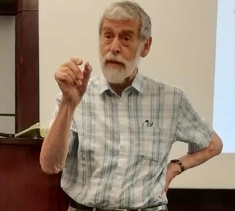
“We’re living at a time when the THz region of the spectrum is now becoming accessible,” said Peter Weightman from the University of Liverpool. “It’s a new tool that we hope may give us advances in cancer management.”
Weightman was a speaker at the recent meeting “Towards the THz Imaging of Cancer“. The event brought together researchers, clinicians and industry players to explore how to transition THz imaging into an effective clinical tool.
Studying single cells
The first speaker, Norbert Klein from Imperial College London, discussed single cell detection based on markers such as cell size or water content, which can be measured using physical techniques. Measurements at THz and microwave frequencies, for example, are sensitive to the water content of a cell, enabling rapid cell characterization without requiring labelling.
“The uniqueness of the microwave to THz range is that it can look inside the cell, but is not yet limited by scattering,” Klein explained. “That’s why this window is of special interest. This is a new form of cancer cell diagnostics that may be complementary to other approaches.”
THz experts: Phil Taday, Norbert Klein and Emma Pickwell-MacPherson.
Klein presented evidence for correlation between microwave response and cancer cell aggressiveness. “We don’t yet know how much we can access with THz, we may see better at THz than microwave frequencies, but this is all early work and more studies are needed,” he added.
Klein and his team developed a cavity-coupled resonator system for fast measurement of flowing cells at 10 GHz (sub-THz). They combined a split-ring resonator with a dielectric resonator, and integrated this into a microfluidic chip. He pointed out that the device is simple to make, low in cost and, in principle, could be extended to 100 GHz (0.1 THz).
They used the device to detect flowing polystyrene microspheres and perform the first microwave measurements of flowing (mouse myoblast) cells. The signals were dependent upon cell volume, providing a fast and accurate way to measure cell size distribution. Potential applications include examining blood samples for circulating tumour cells, which are larger than white blood cells.
For THz detection of single cells, Klein described the use of a silicon photonic crystal resonator to measure blood cell suspensions. “Water content measurement in a single cell is possible,” he concluded. “We have demonstrated this at microwave frequencies and believe it is possible at THz.”
Klein noted that a lab-on-a-chip system that combines rapid measurements of cell size and water content could be a game changer for cancer diagnostics.
In vivo options
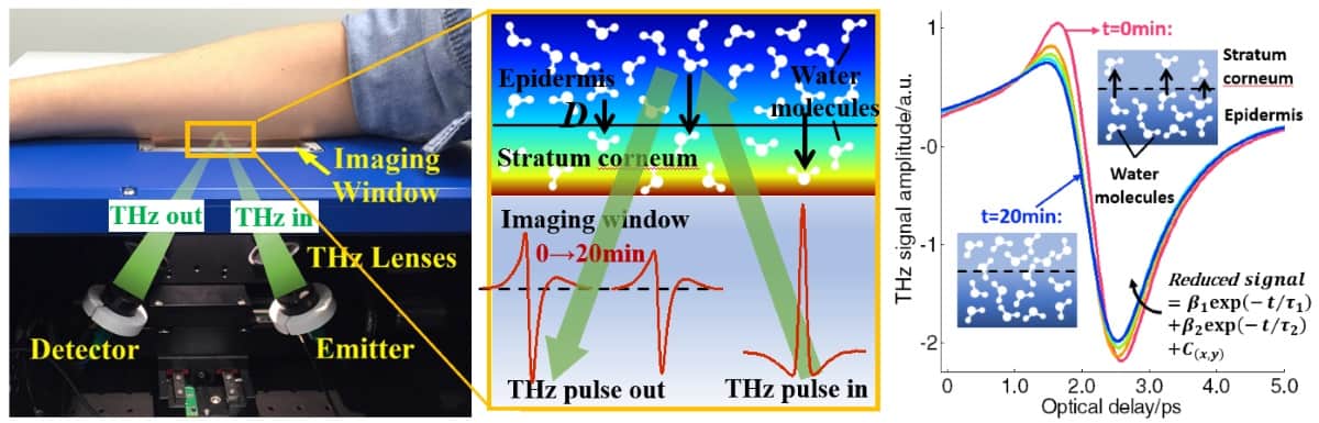
Emma Pickwell-MacPherson from the University of Warwick, examined the challenges of in vivo THz imaging. “We have seen an increase in THz imaging in vivo recently,” she said.
Examples include screening of diabetic foot syndrome, where THz imaging reveals differences in tissue water content between diabetics and controls; monitoring scar healing, in which THz can image subtle tissue changes when visible changes are no longer apparent; and even non-contact THz imaging of the corneal surface.
THz techniques can also detect differences between normal and cancerous tissues. In vivo measurement, however, is challenging, due to the substantial number of variables that need to be controlled. “Measuring the same region of skin each time for comparative studies is tricky,” Pickwell-MacPherson explained. “Skin structure changes with position, pressure, time of day, moisturizer and so on, we need a repeatable protocol.”
In vivo skin measurements are usually performed in reflectance mode, by placing the skin on a quartz window. This, however, occludes the skin, changing its water content and thus decreasing the amplitude of the THz signal, particularly in the first five minutes. “We need to know how to model this effect and compensate for it,” explained Pickwell-MacPherson.
Water molecules accumulate in the stratum corneum during the occlusion process, and the THz response changes accordingly. The skin hydration and water diffusivity can be calculated from the THz data.
Another consideration is variation in the pressure between the skin and the window, which can also affect measurements and required compensation. Pickwell-MacPherson’s team has studied the use of a pressure sensor to monitor such changes.
THz measurements are affected by both the skin’s thickness and its refractive index (hydration). However, the skin hydration also affects its thickness, as skin tends to expand slightly as it hydrates, complicating the analysis of measured signals. One alternative approach is to use ellipsometry, which measures the refractive index independently of thickness.
With this aim, Pickwell-MacPherson and her team have constructed a THz ellipsometer. They noted that the ellipsometry results had much smaller error bars at high frequencies, overcoming some of the difficulties of opaque sample characterization. “Ellipsometry may be ready for in vivo studies soon,” she concluded.
The industry take
Phil Taday of TeraView described the emergence and development of commercial THz instrumentation. TeraView was spun out of Toshiba Research Labs at Cambridge University in 2001, at which time THz systems were the size of optical tables. In 2003, the company launched a smaller, trolley-mounted THz pulsed spectroscopy system, which could differentiate basal cell carcinoma (BCC) from normal tissue samples via absorption and refractive index measurements.
The next development enabled THz imaging, using reflectance mode and a quartz plate. This system could visualize tumour margins, for example to guide surgical excision of skin cancers and ensure all cancer cells are removed.
TeraView has also developed a handheld fibre-coupled THz imaging probe to view excision margins in breast cancer surgery. Here, the THz response could differentiate fat, fibrous tissue and tumour with a sensitivity of 82% and a specificity of more than 92%. A trial of this approach is ongoing at Guy’s Hospital in London.
The company’s latest device is the TeraPulse 4000, which can perform THz imaging and spectroscopy in both transmission and reflection modes. “This is more mobile, we are getting to the stage where the system is now desktop sized,” said Taday.
One future incarnation could be a device for endoscopic THz imaging, for monitoring Barrett’s oesophagus for signs of cancer, for example. Taday said that this should be feasible, noting that THz emitters could be miniaturized to fit into an endoscope channel, but that shrinking the silicon optics used to control the THz beam may perhaps be harder.
High power
In the final presentation, Weightman looked at how THz characterization of tissue could be exploited for cancer diagnosis. He emphasized the need for intense THz sources to perform experiments. “We know that water is very important for biological systems, so if we want to understand the function of DNA in the THz region we have to work in water,” he explained. “The problem is that 1 mm of water attenuates THz by a factor of 1018.”
Current laboratory sources produce THz radiation with intensities of about 100 µW to 10 mW. Energy-recovery linacs, however, generate a peak power of 10 kW, while free electron lasers can generate MW. Such powerful sources allow investigations into how THz radiation impacts the activity, damage and repair of DNA, and whether it changes the expression of genes.
One important question is whether THz radiation is safe. Weightman described a series of experiments performed at the ALICE energy-recovery linac at Daresbury Laboratory, which had a tissue culture facility for THz irradiation of cells within an incubator. “We found that THz radiation had no effect on the morphology, attachment, proliferation or differentiation of several types of cells,” he said.
Weightman noted, however, that another group has shown that intense THz radiation changed the activity of genes in skin cells. He suggested that the live cells used in his studies may have been damaged but then repaired themselves. More experiments are required, and are planned.
With many unanswered questions, Weightman believes it’s essential to perform basic science studies before transitioning to the clinic. “Clinicians want something that they can use in the operating theatre, but at the moment we don’t know what we can tell them,” he explained.
Looking ahead
In panel discussions, the speakers considered the future potential of THz imaging. Pickwell-MacPherson suggested that achieving in vivo THz imaging will require updated instrumentation, pressure sensors and non-contact measurement systems. Klein suggested that it may be optimal to combine infrared, THz and microwave imaging into one device.
“We are still in the process of understanding what THz can tell us,” said Klein. “But once this is found, maybe progression to clinical trials can be quick. If water content and cell size are important, we could be ready in quite a short time for pilot clinical studies. I would encourage to bring clinicians, technologists and physicists to one table.”
Taday emphasized the importance of miniaturization. “That’s down to people like us to drive forward,” he said. There’s also a need to work with academics on areas such as signal processing and how to extract the most from these signals. He added that faster ways to scan the beam are needed, and that array detectors, or THz CCDs, are another key development.
For THz techniques to appeal to the healthcare sector, they need to compete with existing technologies on a real problem, such as cancer management, for example, where anything that could detect early disease is enormously important. Another possibility is rapid assessment of lymph nodes during surgery. And perhaps THz could even be used therapeutically, to preferentially heat and destroy cancer cells.
Weightman pointed out that distinguishing cancer from non-cancer is not difficult, histology can do that. What’s more challenging is to determine whether a tissue will turn cancerous, whether a lesion will progress. “Once you know the target, then you can develop the instruments,” he said.
Participants from the day are taking forward a THz network, to encourage clinicians and THz physicists to develop a coherent strategy for THz-based medical instrumentation and diagnostics. Rob Donnan (r.donnan@qmul.ac.uk) is the point-of-contact for readers interested in joining the network.
Numerous properties are related to the distribution of solvents around solutes, including solvation, free energy, partial molar volume, salting-out constants and binding free energies. However while it is possible to approximate the solvent distribution from rigorous statistical mechanics, determining these properties directly from simple solvent data alone has proved problematic. As Maxim Federov and colleagues point out in their Journal of Physics: Condensed Matter report, “using a purely theoretical approach, it is difficult to relate these [solvent] distributions to the substance’s biological effects which are a result of a large number of complex interrelated phenomena, such as toxicity or bioaccumulation.” Using machine learning based on a 3D convolutional neural network, they show how they can bridge the gap between this simple input data and the complex physical-chemical properties associated with it.
Federov – Director of the Skoltech Center for Computational and Data-Intensive Science and Engineering and a researcher at the Skolkovo Institute of Science and Technology in Estonia and Professor at the University of Strathchlyde in Scotland – worked alongside Sergey Sosnin at Skoltech, Maksim Misin at the University of Tartu in Russia and David S Palmer at the University of Strathclyde. They obtained input data of the concentration of water molecules around various organic molecules from molecular theory using the three-dimensional reference interaction site model. They then split a data set from the USA Environmental Protection Agency into two sets – one to train their neural network and one to test it. The data set comprised measured bioconcentration factor values for various fish species including carp and salmonids.
From simple data to complex properties
Federov and colleagues found that their neural network could determine bioconcentration factors from solvent data with an accuracy equal to the ‘consensus’ model provided by the US EPA. “This result is noteworthy due to the fact that our model was based only on the 3D distribution of water molecules while the EPA’s models used a large set of descriptors of varying nature,” they explain in their report. In contrast results from a graph convolution model were notably less accurate.
The researchers have already simplified their script for the model and further developments are needed to handle the size of the input data to describe the input solvent distribution. However the work demonstrates that artificial neural networks can provide useful links between simple input data and complex physical-chemical properties.
Radioactive molecules have been observed directly in interstellar space for the first time. Astronomers led by Tomasz Kamiński at the Harvard-Smithsonian Center for Astrophysics concluded that aluminium monofluoride containing the unstable isotope aluminium-26 was ejected into space during the merger of binary star system CK Vulpeculae. Their work boosts our understanding of how radioactive aluminium has been dispersed across the Milky Way and could lead to the discovery of other sources of the isotope.
Between 1670 and 1672, astronomers in Europe thought they were witnessing the birth of a new star when a mysterious object called CK Vulpeculae appeared in the sky – emitting red light that was initially bright enough to be seen with the naked eye. We now know that CK Vulpeculae is in the Milky Way about 2300 light-years from Earth, but its precise nature had long puzzled astronomers.
In 2013, the mystery surrounding CK Vulpeculae was partially solved by researchers using the APEX radio telescope in Chile, which spotted a highly unusual molecular gas flowing out from its source. Further analysis suggested that the gas was, in fact, the partial remnant of a rare merger of a low-mass binary star system – which probably produced the light seen in the 1670s.
Gamma-ray sources
When aluminium-26 decays it emits a gamma ray and by detecting this radiation, astronomers have known for several decades that the Milky Way contains about two solar masses of the isotope. However, gamma-ray detectors have poor angular resolution and therefore the sources of aluminium-26 could not be located.
In this latest study, Kamiński’s team used the ALMA radio telescope in Chile, and the NOEMA radio telescope in France to observe the distinctive, millimetre-wavelength signature associated with aluminium monofluoride molecules that contain aluminium-26. This allowed them to conclude that CK Vulpeculae is indeed a source of aluminium-26.
The research provides new insights into how low-mass binary mergers take place – and evidence that heavy elements inside stars can be ejected into space during such mergers.
Other sources needed
The discovery could also give some idea of the origins of the Milky Way’s interstellar aluminium-26. However, the relatively small amount of the isotope emitted by CK Vulpeculae and the current belief that such mergers are exceptionally rare events suggests that there are probably other types of sources of aluminium-26.
The work of Kamiński and colleagues has also shown that modern millimetre-wave interferometers like ALMA could be much better at searching for aluminium-26 than gamma-ray observatories. This could help astronomers look for other sources of the radioactive isotope.
Policies to combat climate change in the short term may not currently be sufficient to realise the long-term goals set in the Paris Agreement. Now researchers in Germany have assessed how to adjust these short-term policies to meet long-term goals while avoiding political and social disruption. Their work could be vital in ensuring the Paris targets are met.
“We show a way how current climate pledges by countries can be strengthened to be more in line with the world’s long-term goal of limiting warming to well below 2 °C,” says Elmar Kriegler at the Potsdam Institute for Climate Research, Germany. “There are political barriers to effective carbon pricing in many countries, so a mix of regulatory policies and moderate carbon pricing can help to keep the long-term goal within reach.”
The researchers calculated that by implementing their proposed regional policies globally, 10 billion tonnes of CO₂ emissions could be avoided by 2030 compared with current plans, greatly reducing the challenges faced by long-term policies.
Though cost-effective, a comprehensive, global set of policies to set a price on carbon emissions is not a realistic prospect in the near future, according to the team. Since the needs of different regions and areas of the economy vary drastically, such all-encompassing measures could quickly result in unrest.
Instead, Kriegler and colleagues suggest a strengthening of existing plans by introducing more moderate policies for different regions, in line with the capabilities of these smaller areas. Such regional policies would include varying degrees of regulation in energy supply, transport, buildings, industry and land use.
“The strengthening of national climate policies and targets until 2030 is instrumental to ensure the success of the Paris Agreement,” Kriegler says. “Thus, national and global conversations on climate change should focus on how to strengthen action, not in the longer-term future, but right now with 2030 in view as a target year.”
Since the UN climate agreement in Paris in December 2015, there have been concerns about how the short-term plans it proposes will only slow, rather than entirely halting, emissions growth in the long term.
Ultimately, this means that actions in the short term may need to ramp up rapidly to reach the agreement’s long-term targets. Such rapid changes could quickly run into political difficulties, including job losses in a variety of sectors due to high carbon prices. Without further measures to combat these short-term policy issues, the goals of the Paris Agreement could be put in jeopardy.
“Short-term climate policies are needed to put the world on a low-carbon pathway,” says Kriegler. “Without stringent short-term policy action, the world’s carbon lock-in will further increase and quickly put the Paris climate goals out of reach. While the climate goals are long-term, our choices today are critical for achieving them.”
In future, Kriegler’s team aims to better describe the global impacts of carbon mitigation policies. Ultimately, the researchers hope to establish a set of indicators to evaluate the scope of climate policies, helping them to create stronger plans for short-term policies.
The “Best-in-Physics” session at the AAPM Annual Meeting showcase the studies that score highest in the abstract review process and are judged to reflect the highest level of scientific quality and innovation. The ever-popular electronic poster session includes presentations in therapy, imaging and joint imaging-therapy categories. Here’s our selection from this year’s chosen papers.
Cardiac compensation enhances CBCT scans
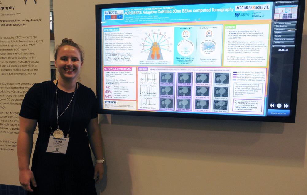
“ACROBEAT is all about improving imaging in the interventional cardiac suite, and eventually within radiotherapy,” explained Tess Reynolds from the University of Sydney. Presenting in the joint imaging-therapy category, she described a 3D gated cardiac cone-beam CT (CBCT) protocol that compensates for cardiac rhythm variations during the scan.
Tess Reynolds from the University of Sydney.
Reynolds explains that, currently, cardiac CBCT scans are not adapted for patient motion. The ACROBEAT (adaptive cardiac cone-beam computed tomography) protocol, on the other hand, uses the patient’s electrocardiograph (ECG) signal to regulate the gantry velocity and projection time intervals in real time.
ACROBEAT decreases the number of projections needed by acquiring all images prospectively in a single gantry rotation. This removes the current need for multiple gantry sweeps and eliminates unnecessary projections not used in image reconstruction.
“This improves the image quality, reduces the dose – which is particularly important for cardiovascular imaging – and significantly reduces scan times,” Reynolds told Physics World. Faster scans help reduce the length of the breath hold required. “We can image adaptively in a single sweep, which gives an 84% reduction in imaging dose.”
To date, Reynolds and colleagues have simulated in silico scans. They are now poised to begin experiments on a research C-arm system, using their software to control the gantry.
Another future aim is monitoring the patient’s breathing trace as well as their ECG to perform dual cardiac- and respiratory-motion mitigated imaging (which could eliminate the need for a breath hold). Such an approach opens up the possibility of using ACROBEAT within radiotherapy, for example to enable targeting of central lung tumours by removing the cardiac motion.
Nanoparticles plus radiation boost drug delivery
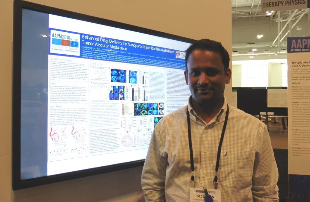
The delivery of cancer drugs to pancreatic tumours is limited by the presence of intact blood vessels that restrict the permeation and penetration of chemotherapeutics or large molecular drugs. Presenting in the therapy category, Sijumon Kunjachan, an instructor in radiation oncology at Harvard Medical School described an innovative technique to address this problem.
Sijumon Kunjachan from Harvard Medical School.
“We are trying to improve drug delivery in pancreatic tumours, by modulating the tumour blood vessels using combined radiation and nanoparticle therapy,” he explained.
The approach is based on the use of gold nanoparticles functionalized with peptides that have an affinity to receptors on tumour endothelial cells. When injected intravenously, the nanoparticles bind to the surface of the tumour blood vessel. During irradiation, these nanoparticles cause the blood vessels to rupture and enable the cancer (nano)drugs to leak through the altered vessels and accumulate in the tumour.
Kunjachan and colleagues tested these nanoparticles in mice bearing a human pancreatic cancer. They injected the functionalized nanoparticles 24 hours prior to external-beam radiation therapy and tested the concept using both preclinical and clinical radiation beams. To determine whether vessel disruption increases permeability, they tested two model drugs: a short-circulating MRI contrast agent and a long-circulating fluorescent contrast.
“We saw that the accumulation of both the agents in pancreatic tumours almost doubled,” said Kunjachan. “The next step will be to test this approach using routinely administered chemotherapeutics or nanodrugs in an orthotopic and genetically engineered pancreatic or lung tumour model, and to observe the tumour regression and survival benefits.”
Selenium ramps TOF-PET performance
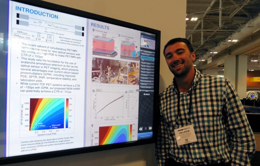
In the imaging category, Andrew LaBella from SUNY Stony Brook University described NEW -HARP, an amorphous selenium photodetector for time-of-flight (TOF) PET. The aim of NEW-HARP (nano-electrode multi-well high-gain avalanche rushing photoconductor) is to achieve a sufficient time resolution to perform TOF-PET in simultaneous PET/MRI.
Andrew LaBella from SUNY Stony Brook University.
LaBella explained that to optimize TOF-PET imaging, the goal is to minimize the coincidence time resolution, ideally to below 100 ps. The resulting improvement in signal-to-noise ratio can then be exploited to increase scanning throughput, reduce dose or enhance lesion detection.
“Current photosensors are costly and have low quantum efficiency,” said LaBella. “So we propose the use of selenium instead of silicon. It is more cost-efficient, offers similar timing and higher quantum efficiency.”
LaBella and colleagues fabricated a multi-well solid-state photomultiplier based on amorphous selenium. To speed up the device performance, they used an embedded grid electrode to achieve unipolar time differential charge sensing – a design that improved the timing resolution to around 100 ps.
The key advantages of NEW-HARP, noted LaBella, are the cost-effectiveness of selenium and the possibility of achieving 100 ps timing. “The benchmark for the field is a coincidence timing resolution of 100 ps, if you can do that, you can justify the cost of TOF-PET and everyone will buy into it,” he explained.
The next steps will be to optimize the fabrication process, and then fabricate and test the coincidence timing of a full ring of PET detectors using NEW-HARP as the photomultiplier coupled to LYSO scintillators.