Nuclear scientists at the Lawrence Berkeley National Laboratory (LBNL) in the US have produced and identified molecules containing nobelium for the first time. This element, which has an atomic number of 102, is the heaviest ever to be observed in a directly-identified molecule, and team leader Jennifer Pore says the knowledge gained from such work could lead to a shake-up at the bottom of the periodic table.
“We compared the chemical properties of nobelium side-by-side to simultaneously produced molecules containing actinium (element number 89),” says Pore, a research scientist at LBNL. “The success of these measurements demonstrates the possibility to further improve our understanding of heavy and superheavy-element chemistry and so ensure that these elements are placed correctly on the periodic table.”
The periodic table currently lists 118 elements. As well as vertical “groups” containing elements with similar properties and horizontal “periods” in which the number of protons (atomic number Z) in the nucleus increases from left to right, these elements are arranged in three blocks. The block that contains actinides such as actinium (Ac) and nobelium (No), as well as the slightly lighter lanthanide series, is often shown offset, below the bottom of the main table.
The end of a predictive periodic table?
Arranging the elements this way is helpful because it gives scientists an intuitive feel for the chemical properties of different elements. It has even made it possible to predict the properties of new elements as they are discovered in nature or, more recently, created in the laboratory.
The problem is that the traditional patterns we’ve come to know and love may start to break down for elements at the bottom of the table, putting an end to the predictive periodic table as we know it. The reason, Pore explains, is that these heavy nuclei have a very large number of protons. In the actinides (Z > 88), for example, the intense charge of these “extra” protons exerts such a strong pull on the inner electrons that relativistic effects come into play, potentially changing the elements’ chemical properties.
“As some of the electrons are sucked towards the centre of the atom, they shield some of the outer electrons from the pull,” Pore explains. “The effect is expected to be even stronger in the superheavy elements, and this is why they might potentially not be in the right place on the periodic table.”
Understanding the full impact of these relativistic effects is difficult because elements heavier than fermium (Z = 100) need to be produced and studied atom by atom. This means resorting to complex equipment such as accelerated ion beams and the FIONA (For the Identification Of Nuclide A) device at LBNL’s 88-Inch Cyclotron Facility.
Producing and directly identifying actinide molecules
The team chose to study Ac and No in part because they represent the extremes of the actinide series. As the first in the series, Ac has no electrons in its 5f shell and is so rare that the crystal structure of an actinium-containing molecule was only determined recently. The chemistry of No, which contains a full complement of 14 electrons in its 5f shell and is the heaviest of the actinides, is even less well known.
In the new work, which is described in Nature, Pore and colleagues produced and directly identified molecular species containing Ac and No ions. To do this, they first had to produce Ac and No. They achieved this by accelerating beams of 48Ca with the 88-Inch Cyclotron and directing them onto targets of 169Tm and 208Pb, respectively. They then used the Berkeley Gas-filled Separator to separate the resulting actinide ions from unreacted beam material and reaction by-products.
The next step was to inject the ions into a chamber in the FIONA spectrometer known as a gas catcher. This chamber was filled with high-purity helium, as well as trace amounts of H2O and N2, at a pressure of approximately 150 torr. After interactions with the helium gas reduced the actinide ions to their 2+ charge state, so-called “coordination compounds” were able to form between the 2+ actinide ions and the H2O and N2 impurities. This compound-formation step took place either in the gas buffer cell itself or as the gas-ion mixture exited the chamber via a 1.3-mm opening and entered a low-pressure (several torr) environment. This transition caused the gas to expand at supersonic speeds, cooling it rapidly and allowing the molecular species to stabilize.
Once the actinide molecules formed, the researchers transferred them to a radio-frequency quadrupole cooler-buncher ion trap. This trap confined the ions for up to 50 ms, during which time they continued to collide with the helium buffer gas, eventually reaching thermal equilibrium. After they had cooled, the molecules were reaccelerated using FIONA’s mass spectrometer and identified according to their mass-to-charge ratio.
A fast and sensitive instrument
FIONA is much faster than previous such instruments and more sensitive. Both properties are important when studying the chemistry of heavy and superheavy elements, which Pore notes are difficult to make, and which decay quickly. “Previous experiments measured the secondary particles made when a molecule with a superheavy element decayed, but they couldn’t identify the exact original chemical species,” she explains. “Most measurements reported a range of possible molecules and were based on assumptions from better-known elements. Our new approach is the first to directly identify the molecules by measuring their masses, removing the need for such assumptions.”
As well as improving our understanding of heavy and superheavy elements, Pore says the new work might also have applications in radioactive isotopes used in medical treatment. For example, the 225Ac isotope shows promise for treating certain metastatic cancers, but it is difficult to make and only available in small quantities, which limits access for clinical trials and treatment. “This means that researchers have had to forgo fundamental chemistry experiments to figure out how to get it into patients,” Pore notes. “But if we could understand such radioactive elements better, we might have an easier time producing the specific molecules needed.”
The way in which new materials are designed is changing, with data becoming ever more important in the discovery and design process. Designing soft materials is a particularly tricky task that requires selection of different “building blocks” (monomers in polymeric materials, for example) and optimization of their arrangement in molecular space.
Soft materials also exhibit many complex behaviours that need to be balanced, and their molecular and structural complexities make it difficult for computational methods to help in the design process – often requiring costly trial and error experimental approaches instead. Now, researchers at Hokkaido University in Japan have combined artificial intelligence (AI) with data mining methods to develop an ultra-sticky hydrogel material suitable for very wet environments – a difficult design challenge because the properties that make materials soft don’t usually promote adhesion. They report their findings in Nature.
Challenges of designing sticky hydrogels
Hydrogels are a permeable soft material composed of interlinked polymer networks with water held within the network. Hydrogels are highly versatile, with properties controlled by altering the chemical makeup and structure of the material.
Designing hydrogels computationally to perform a specific function is difficult, however, because the polymers used to build the hydrogel network can contain a plethora of chemical functional groups, complicating the discovery of suitable polymers and the structural makeup of the hydrogel. The properties of hydrogels are also influenced by factors including the molecular arrangement and intermolecular interactions between molecules (such as van der Waals forces and hydrogen bonds). There are further challenges for adhesive hydrogels in wet environments, as hydrogels will swell in the presence of water, which needs to be factored into the material design.
Data driven methods provide breakthrough
To develop a hydrogel with a strong and lasting underwater adhesion, the researchers mined data from the National Center for Biotechnology Information (NCBI) Protein database. This database contains the amino acid sequences responsible for adhesion in underwater biological systems – such as those found in bacteria, viruses, archaea and eukaryotes. The protein sequences were synthetically mimicked and adapted for the polymer strands in hydrogels.
“We were inspired by nature’s adhesive proteins, but we wanted to go beyond mimicking a few examples. By mining the entire protein database, we aimed to systematically explore new design rules and see how far AI could push the boundaries of underwater adhesion,” says co-lead author Hailong Fan.
The researchers used information from the database to initially design and synthesize 180 bioinspired hydrogels, each with a unique polymer network and all of which showed adhesive properties beyond other hydrogels. To improve them further, the team employed machine learning to create hydrogels demonstrating the strongest underwater adhesive properties to date, with instant and repeatable adhesive strengths exceeding 1 MPa – an order-of-magnitude improvement over previous underwater adhesives. In addition, the AI-designed hydrogels were found to be functional across many different surfaces in both fresh and saline water.
“The key achievement is not just creating a record-breaking underwater adhesive hydrogel but demonstrating a new pathway – moving from biomimetic experience to data-driven, AI-guided material design,” says Fan.
A versatile adhesive
The researchers took the three best performing hydrogels and tested them in different wet environments to show that they could maintain their adhesive properties for long time periods. One hydrogel was used to stick a rubber duck to a rock by the sea, which remained in place despite continuous wave impacts over many tide cycles. A second hydrogel was used to patch up a 20 mm hole on a pipe filled with water and instantly stopped a high-pressure leak. This hydrogel remained in place for five months without issue. The third hydrogel was placed under the skin of mice to demonstrate biocompatibility.
The super strong adhesive properties in wet environments could have far ranging applications, from biomedical engineering (prosthetic coatings or wearable biosensors) to deep-sea exploration and marine farming. The researchers also note that this data-driven approach could be adapted for designing other functional soft materials.
When asked about what’s next for this research, Fan says that “our next step is to study the molecular mechanisms behind these adhesives in more depth, and to expand this data-driven design strategy to other soft materials, such as self-healing and biomedical hydrogels”.
My guest in this episode of the Physics World Weekly podcast is the journalist Jason Palmer, who co-hosts the Intelligence podcast at the Economist.
Palmer did a PhD in chemical physics at Imperial College London before turning his hand to science writing with stints at the BBC and New Scientist.
He explains how he made the transition from the laboratory to the newsroom and offers tips for scientists planning to make the same career journey. We also chat about how artificial intelligence is changing how journalists work.
Traumatic brain injury (TBI), caused by a sudden impact to the head, is a leading cause of death and disability. After such an injury, the most important indicator of how severe the injury is intracranial pressure – the pressure inside the skull. But currently, the only way to assess this is by inserting a pressure sensor into the patient’s brain. UK-based startup Crainio aims to change this by developing a non-invasive method to measure intracranial pressure using a simple optical probe attached to the patient’s forehead.
Can you explain why diagnosing TBI is such an important clinical challenge?
Every three minutes in the UK, someone is admitted to hospital with a head injury, it’s a very common problem. But when someone has a blow to the head, nobody knows how bad it is until they actually reach the hospital. TBI is something that, at the moment, cannot be assessed at the point of injury.
From the time of impact to the time that the patient receives an assessment by a neurosurgical expert is known as the golden hour. And nobody knows what’s happening to the brain during this time – you don’t know how best to manage the patient, whether they have a severe TBI with intracranial pressure rising in the head, or just a concussion or a medium TBI.
Once at the hospital, the neurosurgeons have to assess the patient’s intracranial pressure, to determine whether it is above the threshold that classifies the injury as severe. And to do that, they have to drill a hole in the head – literally – and place an electrical probe into the brain. This really is one of the most invasive non-therapeutic procedures, and you obviously can’t do this to every patient that comes with a blow in the head. It has its risks, there is a risk of haemorrhage or of infection.
Therefore, there’s a need to develop technologies that can measure intracranial pressure more effectively, earlier and in a non-invasive manner. For many years, this was almost like a dream: “How can you access the brain and see if the pressure is rising in the brain, just by placing an optical sensor on the forehead?”
Crainio has now created such a non-invasive sensor; what led to this breakthrough?
The research goes back to 2016, at the Research Centre for Biomedical Engineering at City, University of London (now City St George’s, University of London), when the National Institute for Health Research (NIHR) gave us our first grant to investigate the feasibility of a non-invasive intracranial sensor based on light technologies. We developed a prototype, secured the intellectual property and conducted a feasibility study on TBI patients at the Royal London Hospital, the biggest trauma hospital in the UK.
It was back in 2021, before Crainio was established, that we first discovered that after we shone certain frequencies of light, like near-infrared, into the brain through the forehead, the optical signals coming back – known as the photoplethysmogram, or PPG – contained information about the physiology or the haemodynamics of the brain.
When the pressure in the brain rises, the brain swells up, but it cannot go anywhere because the skull is like concrete. Therefore, the arteries and vessels in the brain are compressed by that pressure. PPG measures changes in blood volume as it pulses through the arteries during the cardiac cycle. If you have a viscoelastic artery that is opening and closing, the volume of blood changes and this is captured by the PPG. Now, if you have an artery that is compromised, pushed down because of pressure in the brain, that viscoelastic property is impacted and that will impact the PPG.
Changes in the PPG signal due to changes arising from compression of the vessels in the brain, can give us information about the intracranial pressure. And we developed algorithms to interrogate this optical signal and machine learning models to estimate intracranial pressure.
How did the establishment of Crainio help to progress the sensor technology?
Following our research within the university, Crainio was set up in 2022. It brought together a team of experts in medical devices and optical sensors to lead the further development and commercialization of this device. And this small team worked tirelessly over the last few years to generate funding to progress the development of the optical sensor technology and bring it to a level that is ready for further clinical trials.
Panicos Kyriacou “At Crainio we want to create a technology that could be used widely, because there is a massive need, but also because it’s affordable.” (Courtesy: Crainio)
In 2023, Crainio was successful with an Innovate UK biomedical catalyst grant, which will enable the company to engage in a clinical feasibility study, optimize the probe technology and further develop the algorithms. The company was later awarded another NIHR grant to move into a validation study.
The interest in this project has been overwhelming. We’ve had a very positive feedback from the neurocritical care community. But we also see a lot of interest from communities where injury to the brain is significant, such as rugby associations, for example.
Could the device be used in the field, at the site of an accident?
While Crainio’s primary focus is to deliver a technology for use in critical care, the system could also be used in ambulances, in helicopters, in transfer patients and beyond. The device is non-invasive, the sensor is just like a sticking plaster on the forehead and the backend is a small box containing all the electronics. In the past few years, working in a research environment, the technology was connected into a laptop computer. But we are now transferring everything into a graphical interface, with a monitor to be able to see the signals and the intracranial pressure values in a portable device.
Following preliminary tests on patients, Crainio is now starting a new clinical trial. What do you hope to achieve with the next measurements?
The first study, a feasibility study on the sensor technology, was done during the time when the project was within the university. The second round is led by Crainio using a more optimized probe. Learning from the technical challenges we had in the first study, we tried to mitigate them with a new probe design. We’ve also learned more about the challenges associated with the acquisition of signals, the type of patients, how long we should monitor.
We are now at the stage where Crainio has redeveloped the sensor and it looks amazing. The technology has received approval by MHRA, the UK regulator, for clinical studies and ethical approvals have been secured. This will be an opportunity to work with the new probe, which has more advanced electronics that enable more detailed acquisition of signals from TBI patients.
We are again partnering with the Royal London Hospital, as well as collaborators from the traumatic brain injury team at Cambridge and we’re expecting to enter clinical trials soon. These are patients admitted into neurocritical trauma units and they all have an invasive intracranial pressure bolt. This will allow us to compare the physiological signal coming from our intracranial pressure sensor with the gold standard.
The signals will be analysed by Crainio’s data science team, with machine learning algorithms used to look at changes in the PPG signal, extract morphological features and build models to develop the technology further. So we’re enriching the study with a more advanced technology, and this should lead to more accurate machine learning models for correctly capturing dynamic changes in intracranial pressure.
The primary motivation of Crainio is to create solutions for healthcare, developing a technology that can help clinicians to diagnose traumatic brain injury effectively, faster, accurately and earlier
This time around, we will also record more information from the patients. We will look at CT scans to see whether scalp density and thickness have an impact. We will also collect data from commercial medical monitors within neurocritical care to see the relation between intracranial pressure and other physiological data acquired in the patients. We aim to expand our knowledge of what happens when a patient’s intracranial pressure rises – what happens to their blood pressures? What happens to other physiological measurements?
How far away is the system from being used as a standard clinical tool?
Crainio is very ambitious. We’re hoping that within the next couple of years we will progress adequately in order to achieve CE marking and all meet the standards that are necessary to launch a medical device.
The primary motivation of Crainio is to create solutions for healthcare, developing a technology that can help clinicians to diagnose TBI effectively, faster, accurately and earlier. This can only yield better outcomes and improve patients’ quality-of-life.
Of course, as a company we’re interested in being successful commercially. But the ambition here is, first of all, to keep the cost affordable. We live in a world where medical technologies need to be affordable, not only for Western nations, but for nations that cannot afford state-of-the-art technologies. So this is another of Crainio’s primary aims, to create a technology that could be used widely, because there is a massive need, but also because it’s affordable.
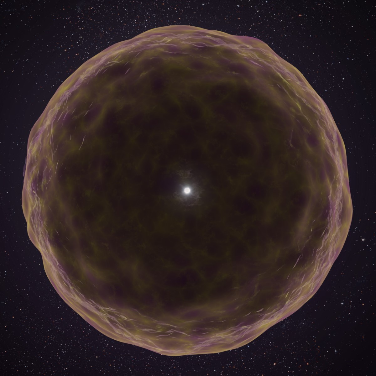
Stripped star Artist’s impression of the star that exploded to create SN 2021yfj. Shown are the ejection of silicon (grey), sulphur (yellow) and argon (purple) just before the final explosion. (Courtesy: WM Keck Observatory/Adam Makarenko)
For the first time, astronomers have observed clear evidence for a heavily stripped star that has shed many of its outer layers before its death in a supernova explosion. Led by Steve Schulze at Northwestern University, the team has spotted the spectral signatures of heavier elements that are usually hidden deep within stellar interiors.
Inside a star, atomic nuclei fuse together to form heavier elements in a process called nucleosynthesis. This releases a vast amount of energy that offsets the crushing force of gravity.
As stars age, different elements are consumed and produced. “Observations and models of stars tell us that stars are enormous balls of hydrogen when they are born,” Schulze explains. “The temperature and density at the core are so high that hydrogen is fused into helium. Subsequently, helium fuses into carbon, and this process continues until iron is produced.”
Ageing stars are believed to have an onion-like structure, with a hydrogen outer shell enveloping deeper layers of successively heavier elements. Near the end of a star’s life, inner-shell elements including silicon, sulphur, and argon fuse to form a core of iron. Unlike lighter elements, iron does not release energy as it fuses, but instead consumes energy from its surroundings. As a result, the star can no longer withstand its own gravity and it will collapse rapidly in and then explode in a dramatic supernova.
Hidden elements
Rarely, astronomers can observe an old star that has blown out its outer layers before exploding. When the explosion finally occurs, heavier elements that are usually hidden within deeper shells create absorption lines in the supernova’s light spectrum, allowing astronomers to determine the compositions of these inner layers. So far, inner-layer elements as heavy as carbon and oxygen have been observed, but not direct evidence for elements in deeper layers.
Yet in 2021, a mysterious new observation was made by a programme of the Zwicky Transient Facility headed by Avishay Gal-Yam at the Weizmann Institute of Science in Israel. The team was scanning the sky for signs of infant supernovae at the very earliest stages following their initial explosion.
“On 7 September 2021 it was my duty to look for infant supernovae,” Schulze recounts. “We discovered SN 2021yfj due to its rapid increase in brightness. We immediately contacted Alex Filippenko’s group at the University of California Berkeley to ask whether they could obtain a spectrum of this supernova.”
When the results arrived, the team realised that the absorption lines in the supernova’s spectrum were unlike anything they had encountered previously. “We initially had no idea that most of the features in the spectrum were produced by silicon, sulphur, and argon,” Schulze continues. Gal-Yam took up the challenge of identifying the mysterious features in the spectrum.
Shortly before death
In the meantime, the researchers examined simultaneous observations of SN 2021yfj, made by a variety of ground- and space-based telescopes. When Gal-Yam’s analysis was complete, all of the team’s data confirmed the same result. “We had detected a supernova embedded in a shell of material rich in silicon, sulphur, and argon,” Schulze describes. “These elements are formed only shortly before a star dies, and are often hidden beneath other materials – therefore, they are inaccessible under normal circumstances.”
The result provided clear evidence that the star had been more heavily stripped back towards the end of its life than any other observed previously: shedding many of its outer layers before the final explosion.
“SN 2021yfj demonstrates that stars can die in far more extreme ways than previously imagined,” says Schulze. “It reveals that our understanding of how stars evolve and die is still not complete, despite billions of them having already been studied.” By studying their results, the team now hopes that astronomers can better understand the later stages of stellar evolution, and the processes leading up to these dramatic ends.
Rainer Weiss, who shared the Nobel Prize for Physics in 2017 for the discovery of gravitational waves, died on 25 August at the age of 92. Weiss came up with the idea of detecting gravitational waves by measuring changes in distance as tiny as 10–18 m via an interferometer several kilometres long. His proposal eventually led to the formation of the twin Laser Interferometer Gravitational-Wave Observatory (LIGO), which first detected such waves in 2015.
Weiss was born in Berlin, Germany, on 29 September 1932 shortly before the Nazis rose to power. With a father who was Jewish and an ardent communist, Weiss and his family were forced to flee the country – first to Czechoslovakia and then to the US in 1939. Weiss was raised in New York, finishing his school days at the private Columbia Grammar School thanks to a scholarship from a refugee relief organization.
In 1950 Weiss began studying electrical engineering at Massachusetts Institute of Technology (MIT) before switching to physics, eventually earning a PhD in 1962, developing atomic clocks under the supervision of Jerrold Zacharias,. He then worked at Tufts University before moving to Princeton University, where he was a research associate with the astronomer and physicist Robert Dicke.
In 1964 Weiss returned to MIT, where he began developing his idea of using a large interferometer to measure gravitational waves. Teaming up with Kip Thorne at the California Institute of Technology (Caltech), Weiss drew up a feasibility study for a kilometre-scale laser interferometer. In 1979 the National Science Foundation funded Caltech and MIT to develop the proposal to build LIGO.
Construction of two LIGO detectors – one in Hanford, Washington and the other at Livingston, Louisiana, each of which featured arms 4 km long – began in 1990, with the facilities opening in 2002. After almost a decade of operation, however, no waves had been detected so in 2011 the two observatories were upgraded to make them 10 times more sensitive than before.
On 14 September 2015 – during the first observation run of what was known as Advanced LIGO, or aLIGO – the interferometer detected gravitational waves from two merging black holes some 1.3 billion light-years from Earth. The discovery was announced by those working on aLIGO in February 2016.
The following year, Weiss was awarded one half of the 2017 Nobel Prize for Physics “for decisive contributions to the LIGO detector and the observation of gravitational waves”. The other half was shared by Thorne and fellow Caltech physicist Barry Barish, who was LIGO project director.
‘An indelible mark’
As well as pioneering the detection of gravitational waves, Weiss also developed atomic clocks and led efforts to measure the spectrum of the cosmic microwave background via weather balloons. He co-founded NASA’s Cosmic Background Explorer project, measurements from which have helped support the Big Bang theory describing the expansion of the universe.
In addition to the Nobel prize, Weiss was awarded the Gruber Prize in Cosmology in 2006, the Einstein Prize from the American Physical Society in 2007 as well as the Shaw Prize and the Kavli Prize in Astrophysics, both in 2016.
MIT’s dean of science Nergis Mavalvala, who worked with Weiss to build an early prototype of a gravitational-wave detector as part of her PhD in the 1990s, says that every gravitational-wave event that is observed “will be a reminder of his legacy”.
“[Weiss] leaves an indelible mark on science and a gaping hole in our lives,” says Mavalvala. “I am heartbroken, but also so grateful for having him in my life, and for the incredible gifts he has given us – of passion for science and discovery, but most of all to always put people first.”
Scientists at the Massachusetts Institute of Technology (MIT) in the US have achieved the cleanest demonstration yet of the famous double-slit experiment. Using two single atoms as the slits, they inferred the photon’s path by measuring subtle changes in the atoms’ properties after photon scattering. Their results matched the predictions of quantum theory: interference fringes when no path was observed, two bright spots when it was.
First performed in the 1800s by Thomas Young, the double-slit experiment has been revisited many times. Its setup is simple: send light toward a pair of slits in a screen and watch what happens. Its outcome, however, is anything but. If the light passes through the slits unobserved, as it did in Young’s original experiment, an interference pattern of bright and dark fringes appears, like ripples overlapping in a pond. But if you observe which slit the light goes through, as Albert Einsten proposed in a 1920s “thought experiment” and as other physicists have since demonstrated in the laboratory, the fringes vanish in favour of two bright spots. Hence, whether light acts as a wave (fringes) or a particle (spots) depends on whether anyone observes it. Reality itself seems to shift with the act of looking.
The great Einstein–Bohr debate
Einstein disliked the implications of this, and he and Niels Bohr debated them extensively. According to Einstein, observation only has an effect because it introduces noise. If the slits were mounted on springs, he suggested, their recoil would reveal the photon’s path without destroying the fringes.
Bohr countered that measuring the photon’s recoil precisely enough to reveal its path would blur the slits’ positions and erase interference. For him, this was not a flaw of technology but a law of nature – namely, his own principle of complementarity, which states that quantum systems can show wave-like or particle-like behaviour, but never both at once.
Physicists have performed numerous versions of the experiment since, and each time the results have sided with Bohr. Yet the unavoidable noise in real set-ups left room for doubt that this counterintuitive rule was truly fundamental.
Atoms as slits
To celebrate the International Year of Quantum Science and Technology, physicists in Wolfgang Ketterle’s group at MIT performed Einstein’s thought experiment directly. They began by cooling more than 10,000 rubidium atoms to near absolute zero and trapping them in a laser-made lattice such that each one acted as an individual scatterer of light. If a faint beam of light was sent through this lattice, a single photon could scatter off an atom.
Since the beam was so faint, the team could collect very little information per experimental cycle. “This was the most difficult part,” says team member Hanzhen Lin, a PhD student at MIT. “We had to repeat the experiment thousands of times to collect enough data.”
In every such experiment, the key was to control how much photon path information the atoms provided. The team did this by adjusting the laser traps to tune the “fuzziness” of the atoms’ position. Tightly trapped atoms had well-defined positions and so, according to Heisenberg’s uncertainty principle, they could not reveal much about the photon’s path. In these experiments, fringes appeared. Loosely trapped atoms, in contrast, had more position uncertainty and were able to move, meaning an atom struck by a photon could carry a trace of that interaction. This faint record was enough to collapse the interference fringes, leaving only spots. Once again, Bohr was right.
While Lin acknowledges that theirs is not the first experiment to measure scattered light from trapped atoms, he says it is the first to repeat the measurements after the traps were removed, while the atoms floated freely. This went further than Einstein’s spring-mounted slit idea, and (since the results did not change) eliminated the possibility that the traps were interfering with the observation.
“I think this is a beautiful experiment and a testament to how far our experimental control has come,” says Thomas Hird, a physicist who studies atom-light interactions at the University of Birmingham, UK, and was not involved in the research. “This probably far surpasses what Einstein could have imagined possible.”
The MIT team now wants to observe what happens when there are two atoms per site in the lattice instead of one. “The interactions between the atoms at each site may give us interesting results,” Lin says.
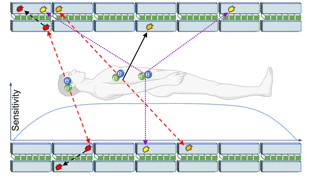
Positron emission tomography (PET) is a diagnostic imaging technique that uses an injected radioactive tracer to detect early signs of cancer, brain disorders or other diseases. At Jagiellonian University in Poland, a research team headed up by Paweł Moskal is developing a totally new type of PET scanner. The Jagiellonian PET (J-PET) can image the properties of positronium, a positron–electron bound state produced during PET scans, offering potential to increase the specificity of PET diagnoses.
The researchers have now recorded the first ever in vivo positronium image of the human brain. They also used the J-PET to show that annihilation photons generated during PET scans are not completely quantum entangled, opening up the possibility of using the degree of quantum entanglement as a diagnostic indicator. Moskal tells Physics World’s Tami Freeman about these latest breakthroughs and the team’s ongoing project to build the world’s first whole-body quantum PET scanner.
Can you describe how conventional PET images are generated?
PET is based on the annihilation of a positron with an electron to create two photons. The patient is administered a radiopharmaceutical labelled with a positron-emitting radionuclide (for example, fluoro-deoxy-glucose (FDG) labelled with 18F), which localizes in targeted tissues. The 18F emits positrons inside the body, which annihilate with electrons from the body, and the resulting annihilation photons are registered by the PET scanner.
By measuring the locations and times of the photons’ interactions in the scanner, we can reconstruct the density distribution of annihilation points in the body. With 18F-FDG, this image correlates with the density distribution of glucose, which in turn, indicates the rate of glucose metabolism. Thus the PET scanner delivers an image of the radiopharmaceutical’s metabolic rate in the body.
Such an image enables physicians to identify tissues with abnormal metabolism, such as cancers that metabolize glucose up to 10 times more intensively than healthy tissues. Therefore, PET scanners can provide information about alterations in cell function, even before cancer may be visible in anatomical images recorded using CT or MRI.
During annihilation, a short-lived atom called positronium can form. What’s the rationale for imaging this positronium?
It’s amazing that in tissue, positron–electron annihilation proceeds via the formation of positronium in about 40% of cases. Positronium, a bound state of matter and antimatter (an electron and a positron), is short lived because it can undergo self-annihilation into photons. In tissue, however, it can decay via additional processes that further shorten its lifetime. For example, its positron may annihilate by “picking off” an electron from a surrounding atom, or it may convert from the long-lived state (ortho-positronium) to the short-lived state (para-positronium) through interaction with oxygen molecules.
In tissue, therefore, positronium lifetime is an indicator of the intra- and inter-molecular structure and the concentration of oxygen molecules. Both molecular composition and the degree of oxygen concentration differ between healthy and cancerous tissues, with hypoxia (a deficit in tissue oxygenation) a major feature of solid tumours that’s related to the development of metastases and treatment resistance.
As such, imaging positronium lifetime can help in early disease recognition at the stage of molecular alterations. It can also improve diagnosis and the proper choice of anti-cancer therapy. In the case of brain diagnostics, positronium imaging may become an early diagnostic indicator for neurodegenerative disorders such as dementia, Alzheimer’s disease and Parkinson’s disease.
So how does the J-PET detect positronium?
To reconstruct the positronium lifetime we use a radionuclide (44Sc, 82Rb or 124I, for example) that, after emitting a positron, promptly (within a few picoseconds) emits an additional gamma photon. This “prompt gamma” can be used to measure the exact time that the positron was emitted into the tissue and formed positronium.
Multiphoton detection In about 1% of cases, after emitting a positron that annihilates with an electron into photons (blue arrows), 68Ga also emits a prompt gamma (solid arrow). (Courtesy: CC BY/Sci. Adv. 10.1126/sciadv.adp2840)
Current PET scanners are designed to register only two annihilation photons, which makes them incapable of determining positronium lifetime. The J-PET is the first multiphoton PET scanner designed for simultaneous registration of any number of photons.
The registration of annihilation photons enables us to reconstruct the time and location of the positronium decay, while registration of the prompt gamma provides the time of its formation. The positronium lifetime is then calculated as the time difference between annihilation and prompt gamma emission.
Can you describe how your team recorded the first in vivo positronium image?
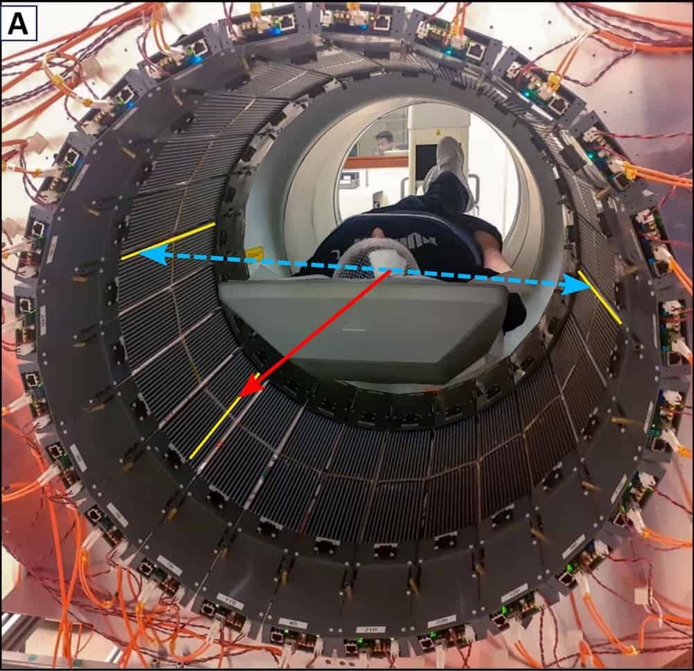
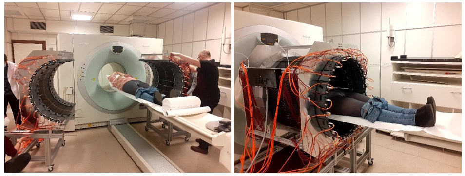
Last year we presented the world’s first in vivo images of positronium lifetime in a human, reported in Science Advances. For this, we designed and constructed a modular, lightweight and portable J-PET tomograph, consisting of 24 independent detection modules, each weighing only 2 kg. The device uses a multiphoton data acquisition system, invented by us, to simultaneously register prompt gamma and annihilation photons – the first PET scanner in the world to achieve this.
The research was performed at the Medical University of Warsaw, with studies conducted following routine procedures so as not to interfere with routine diagnostics and therapy. If a patient agreed to stay longer on the platform, we had about 10 minutes to install the J-PET tomograph around them and collect data.
In vivo imaging The first imaging of a patient, illustrating the advantages of the J-PET as a portable, lightweight device with an adaptable imaging volume. (Courtesy: Paweł Moskal)
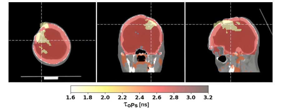
The first patient was a 45-year-old man with glioblastoma (an aggressive brain tumour) undergoing alpha-particle radiotherapy. The primary aim of his therapy was to destroy the tumour using alpha particles emitted by the radionuclide 225Ac. The positronium imaging was made possible by the concurrent theranostic application of the radionuclide 68Ga to monitor the site of cancer lesions using a PET scanner.
The patient was administered a simultaneous intra-tumoural injection of the alpha-particle-emitting radiopharmaceutical (225Ac-DOTA-SP) for therapy and the positron emitting pharmaceutical (68Ga-DOTA-SP) for diagnosis. In about 1% of cases, after emitting a positron that annihilates with an electron, 68Ga also emits a prompt gamma ray.
We determined the annihilation location by measuring the time and position of interaction of the annihilation photons in the scanner. For each image voxel, we also determined a lifetime spectrum as the distribution of differences between the time of annihilation and the time of prompt gamma emission.
Our study found that positronium lifetimes in glioblastoma cells are shorter than in salivary glands and healthy brain tissues. We showed for the first time that the mean lifetime of ortho-positronium in a glioma (1.77±0.58 ns) is shorter than in healthy brain tissue (2.72±0.72 ns). This finding demonstrates that positronium imaging could be used for in vivo diagnosis to differentiate between healthy and cancerous tissues.
Lifetime distributions Positronium images of a patient with glioblastoma, showing the difference in mean ortho-positronium lifetime between glioma and healthy brain. (Courtesy: CC BY/Sci. Adv. 10.1126/sciadv.adp2840)
You recently demonstrated that J-PET can detect quantum entanglement of annihilation photons, how could this impact cancer diagnostics?
For this study, reported earlier this year in Science Advances, we used the laboratory prototype of the J-PET scanner (as employed previously for the first ex vivo positronium imaging). The crucial result was the first ever observation that photons from electron–positron annihilation in matter are not completely quantum entangled. Our study is pioneering in revealing a clear dependence of the degree of photon entanglement on the material in which the annihilation occurs.
These results are totally new compared with all previous investigations of photons from positron–electron annihilations. Up to this point, all experiments had focused on showing that this entanglement is maximal, and for that purpose, were performed in metals. None of the previous studies mentioned or even hypothesized a possible material dependence.
Lab prototype The J-PET scanner used to discover non-maximal entanglement, with (left to right) Deepak Kumar, Sushil Sharma and Pawel Moskal. (Courtesy: Damian Gil and Deepak Kumar)
If the degree of quantum entanglement of annihilation photons depends on the material, it may also differ according to tissue type or the degree of hypoxia. This is a hypothesis that we will test in future studies. I recently received an ERC Advanced Grant, entitled “Can tissue oxidation be sensed by positronium?”, to investigate whether the degree of oxidation in tissue can be sensed by the degree of quantum entanglement of photons originating from positron annihilation.
What causes annihilation photons to be entangled (or not)?
Quantum entanglement is a fascinating phenomenon that cannot be explained by our classical perception of the world. Entangled photons behave as if one instantly knows what is happening with the other, regardless of how far apart they are, so they propagate in space as a single object.
Annihilation photons are entangled if they originate from a pure quantum state. A state is “pure” if we know everything that can be known about it. For example, if the photons originate from the ground state of para-positronium (a pure state), then we expect them to be maximally entangled.
However, if electron–positron annihilation occurs in a mixed state (a statistical mixture of different pure states) where we have incomplete information, then the resulting photons will not be maximally entangled. In our case, this could be the annihilation of a positron from positronium with electrons from the patient’s body. Because these electrons can have different angular momenta with respect to the positron, the annihilation generally occurs from a mixed state.
You have also measured the polarization of the annihilation photons; how is this information used?
In current PET scanners, images are reconstructed based on the position and time of interaction of annihilation photons within the scanner. However, annihilation photons also carry information about their polarization.
Theoretically, annihilation photons are quantum entangled in polarization and exhibit non-local correlations. In the case of electron–positron annihilation into two photons, this means that the amplitude of the distribution of the relative angle between their polarization planes is larger when they are quantum entangled than when they propagate in space as independent objects.
State-of-the-art PET scanners, however, cannot access polarization information. Annihilation photons have energy in the mega-electronvolt range and their polarization cannot be determined using established optical methods, which are designed for optical photons in the electronvolt range. Because these energetic annihilation photons interact with single electrons, their polarization can only be sensed via Compton scattering.
The angular distribution of photons scattered by electrons is not isotropic with respect to the polarization direction. Instead, scattering is most likely to occur in a plane perpendicular to the polarization plane of the photon before scattering. Thus, by determining the scattering plane (containing the primary and scattered photon), one can estimate the direction of polarization as being perpendicular to that plane. Therefore, to practically determine the polarization plane of the photon, you need to know its directions of flight both before and after Compton scattering in the material.
In plastic scintillators, annihilation photons primarily interact via the Compton effect. As the J-PET is built from plastic scintillators, it’s ideally suited to provide information about the photons’ polarization, which can be determined by registering both the annihilation photon and the scattered photon and then reconstructing the scattering plane.
Using the J-PET scanner, we determined the distribution of the relative angle between the polarization planes of photons from positron–electron annihilation in a porous polymer. The amplitude of the observed distribution is smaller than predicted for maximally quantum-entangled two-photon states, but larger than expected for separable photons.
This result can be explained by assuming that photons from pick-off annihilation are not entangled, while photons from direct and para-positronium annihilations are maximally entangled. Our finding indicates that the degree of entanglement depends on the annihilation mechanism in matter, opening avenues for exploring polarization correlations in PET as a diagnostic indicator.
What further developments are planned for the J-PET scanner?
When creating the J-PET technology, we started with a two-strip prototype, then a 24-strip prototype in 2014, followed by a full-scale 192-strip prototype in 2016. In 2021 we completed the construction of a lightweight (60 kg) J-PET version that is both modular and portable, and which we used to demonstrate the first clinical images.
The next step is the construction of the total-body quantum J-PET scanner. We are now at the stage of collecting all the elements of this scanner and expect to complete construction in 2028. The scanner will be installed at the Center for Theranostics, established by myself and Ewa Stępień, medical head of the J-PET team, at Jagiellonian University.
Future developments Schematic cross-section of the full-body J-PET scanner under construction at Jagiellonian University. The diagram shows the patient and several examples of electron–positron annihilation. (Courtesy: Rev. Mod. Phys. 10.1103/RevModPhys.95.021002)
Total-body PET provides the ability to image the metabolism of all tissues in the body at the same time. Additionally, due to the high sensitivity of total-body PET scanners, it is possible to perform dynamic imaging – essentially, creating a movie of how the radiopharmaceutical distributes throughout the body over time.
The total-body J-PET will also be able to register the pharmacokinetics of drugs administered to a patient. However, its true distinction is that it will be the world’s first quantum PET scanner with the ability to image the degree of quantum entanglement of annihilation photons throughout the patient’s body. Additionally, it will be the world’s first total-body multiphoton PET, enabling simultaneous positronium imaging in the entire human body.
How do you see the J-PET’s clinical applications evolving in the future?
We have already performed the first clinical imaging using J-PET at the Medical University of Warsaw and the University Hospital in Kraków. The studies included the diagnosis of patients with neuroendocrine, prostate and glioblastoma tumours. The data collected at these hospitals were used to reconstruct standard PET images as well as positronium lifetime images.
Next, we plan to conduct positronium imaging of phantoms and humans with various radionuclides to explore its clinical applications as a biomarker for tissue pathology and hypoxia. We also intend to explore the J-PET’s multiphoton capabilities for simultaneous double-tracer imaging, as well as study the degree of quantum entanglement as a function of the annihilation mechanism.
Finally, we plan to explore the possibilities of applying quantum entanglement to diagnostics, and we look forward to performing total-body positronium and quantum entanglement imaging with the total-body J-PET in the Center for Theranostics.
Paweł Moskal is a panellist in the forthcoming Physics World Live event on 25 September 2025. The event, which also features Miles Padgett from the University of Glasgow and Matt Brookes from the University of Nottingham, will examine how medical physics can make the most of the burgeoning field of quantum science. You can sign up free here.
Imagine a molecule moving randomly through a cell. Its goal is to bind to an enzyme, a process essential for many chemical reactions in the body.
However, the enzyme isn’t always ready to bind. It switches between two states: a reactive state, where it can bind the molecule, and a non-reactive state, where it cannot.
Even if the molecule reaches the enzyme, binding will only take place if the enzyme happens to be in its reactive state at that exact moment. Simply arriving at the enzyme isn’t enough -the gate must also be open.
This scenario is an example of a gated first-passage process. The term refers to situations where an event (like binding) only happens if two conditions are met: the particle must reach a specific target, and the target must be in the right state to allow the event to occur.
These processes are important in a wide range of fields including chemistry, biophysics, finance, and climate science.
Existing models have offered valuable insights in simple cases, such as point targets, but even those have left some questions unresolved — and the challenges only deepen in more realistic scenarios involving extended targets or thresholds.
In order to address this problem, a team of researchers from India and Israel set about developing a new mathematical framework to analyse these type of processes.
The new approach uses a concept called renewal theory to break down complex processes into simpler, repeatable parts. Renewal theory is a branch of probability that deals with events that happen repeatedly over time, with random intervals between them.
The team showed that their method can solve previously unsolved problems and reveals universal patterns in how long these processes take. They were even able to explain surprising effects that previously posed a major challenge.
Crucially, their method can be applied to many real-world systems, from chemical reactions to intermittent data monitoring.
Interesting phenomena in quantum materials are often found near boundaries between different competing ground states.
Understanding the competition between these states is a central problem in condensed matter physics because of the potential applications to quantum computing and superconductivity.
There are many different types of ground states but the one that’s important here is a charge density wave (CDW). This is where the electron density of a material becomes modulated in a periodic pattern.
TbTe₃, or terbium tritelluride is a quasi-two-dimensional material made up of alternating layers of conducting tellurium (Te) planes and insulating rare-earth terbium (Tb) block layers.
It has attracted a lot of interest recently though because it has two competing CDW states and represents an excellent platform to study new quantum phenomena.
Previous experiments have shown that these states can be tuned when the material is put under pressure, even leading to an induced superconducting state.
These experiments all used an isotropic pressure – the same in all directions. However, because this material is quasi-two dimensional, it would be even more interesting to see how it responds to a strain in one particular direction.
This is exactly what the team at SLAC have done.
They used ultrafast optical reflectivity to probe the dynamics of the competing CDW states in TbTe₃ at different strains.
They found that these two competing states are incredibly similar in energy and become more stable with increasing strain.
What’s really exciting though is the method they used. Their measurements were recorded in a pump-probe setup on timescales of a couple of picoseconds (trillionths of a second).
Combined with the application of a directional strain, this technique could be used in the future to study many other quantum materials with exciting properties.