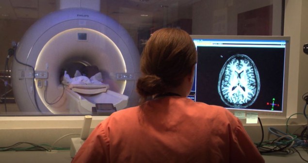
A research team headed up at Keck School of Medicine of USC is using ultrahigh-field MRI to investigate the relationship between migraine and microvascular changes in the brain. The researchers have identified, for the first time, that migraine sufferers exhibit enlarged perivascular spaces – fluid-filled spaces surrounding blood vessels – in their brains. Study co-author Wilson Xu reported their findings at this week’s RSNA 2022, the annual meeting of the Radiological Society of North America.
Migraine is a common condition characterized by a severe recurring headache, often accompanied by nausea, weakness and light sensitivity. Xu and colleagues used 7T MRI to study microvascular changes in the brain due to different types of migraine. They note that because ultrahigh-field MRI can create images with higher resolution and better quality than other types of MR scan, it can reveal brain tissue changes after a migraine in more detail.
“In people with chronic migraine and episodic migraine without aura, there are significant changes in the perivascular spaces of a brain region called the centrum semiovale. These changes have never been reported before,” explains Xu. “Perivascular spaces are part of a fluid clearance system in the brain. Studying how they contribute to migraine could help us better understand the complexities of how migraines occur.”
The researchers performed a series of 7T MRI exams – T1, T2, FLAIR and SWI/QSM sequences – on 10 participants with chronic migraine, 10 with episodic migraine without aura and five age-matched healthy controls. They used the MR data to calculate the size of perivascular spaces in the centrum semiovale (the central area of white matter) and basal ganglia (a group of structures primarily responsible for motor control) of the brain.
Preliminary statistical analysis revealed that the number of enlarged perivascular spaces in the centrum semiovale, but not in the basal ganglia, was significantly higher in patients with either type of migraine than in healthy controls.
The team also measured cerebral microbleeds and white matter hyperintensities – lesions in the brain that appear as areas of increased brightness on MRI. There were no significant differences in the frequency of microbleeds or hyperintensities between patients with or without migraine. Migraine patients, however, showed a significant correlation between the presence of enlarged perivascular spaces and the severity of white matter lesions.

Exploring the visual symptoms of migraine
The researchers plan to continue their study with larger populations and ongoing follow-up, with the aim of better understanding the relationship between structural changes and migraine development and type.
“The results of our study could help inspire future, larger-scale studies to continue investigating how changes in the brain’s microscopic vessels and blood supply contribute to different migraine types,” Xu says. “Eventually, this could help us develop new, personalized ways to diagnose and treat migraine.”


