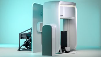
Interpreting images that might contain tumours can be difficult. Biological tissue contains a variety of fluids and other material that increase the chances of registering 'ghost' images, or hide the tumour amongst the 'noise'. Now Sel Colak has developed a mathematical method to improve such images.
The technique works on any turbid medium observed with light in the wavelength range 200-1000 nm, making it suitable for a wide range of applications. Patent 5719398 describes how three simple stages create the final image.
First the observer measures the optical parameters of the object and convolutes the data with a special filter. A light distribution function is then applied to model the passage of light through the medium. A final step cleans the image of any high-frequency noise by using a low-pass filter. The main advantage of this system is that fewer light sources are required to produce high-quality images, but high-quality real-time images are unavailable with the technique at present.


