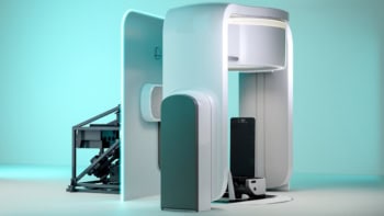
A human heartbeat has been imaged by a magnetic resonance scanner for the first time. Greg Hundley and colleagues at the Wake Forest University Baptist Medical Center in North Carolina have developed a software program that can analyse a magnetic resonance scanner within seconds, rather than the five minutes needed at present. This allows doctors to look at images of a beating heart in close to real-time. The team hopes to use the technique to look for abnormal pumping motions that could indicate blocked arteries (Circulation 100 1697).
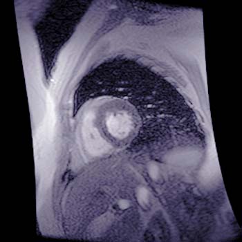
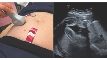
Ultrasound is commonly used to look at the heart, but it is unsuitable for the 10-20% of the population that have breathing difficulties (emphysema) or who are medically obese. In both techniques, patients are given drugs to speed up their hearts and promote vigorous pumping. In a healthy heart both sides of the heart contract with equal force. If some of the blood vessels are blocked, however, one side of the heart wall will not contract normally.
Magnetic resonance imaging also allows the patient to be checked for symptoms of other medical conditions. “MRI allows us not only to locate a blockage, but to determine whether it limits blood flow enough to warrant treatment,” says Hundley. “A lot of new doors have been opened with this technology.”
