A new medical imaging technique could help doctors to detect osteoporosis and other conditions related to weak bones in old people. Qingwen Ni and colleagues at the Southwest Research Institute and the University of Texas have used nuclear magnetic resonance (NMR) to measure the porosity of bone. Increases in porosity cause bones to become weaker, thus leading to a greater risk of fracture (Q Ni et al. 2004 Meas. Sci. Technol. 15 58)

NMR is already widely used to measure porosity in materials such as rocks, concrete and wood. Magnetic pulses are first used to align the spins of certain nuclei in the sample. The nuclei then relax and emit ‘echoes’ – lasting up to seconds – which show how abundant that particular nucleus is in the sample.
Ni and colleagues have now used a low field (0.5 Tesla) pulsed NMR to analyse the relaxation time of hydrogen nuclei (protons) in samples of bone taken from donors aged 19 to 89 years. These protons are present in the water-like fluid inside the pores of bone. The relaxation time is proportional to the amount of liquid inside the pores and thus the porosity of the bone. Moreover, the data can also provide information about the distribution of pore sizes.
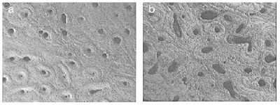
The researchers found that the average porosity was about 8.5% for samples less than 45 years old, and about 17% for those over 63. The average pore size in older bone was also larger – between 50 and 100 microns, compared with 10-50 microns for younger samples (see micrographs). The results agree with measurement made with routinely used – but destructive – methods. “Since this NMR technique is non-invasive and non-destructive, it has great potential in the biomedical fields, particularly in bone-related research and applications,” says Ni.


