Claustrophobic patients who fear lying inside the tight confines of a conventional magnetic resonance imaging (MRI) scanner could have help at hand. Physicists in the US have created a new MRI device that patients can literally walk into and be studied standing up or sitting down. It uses much lower magnetic fields than conventional scanners and is designed to study patients who suffer from lung disease (R W Mair et al. 2004 arXiv.org/abs/physics/0403090). The only snag to the device, which has been created by a team led by Ronald Walsworth at the Harvard-Smithsonian Center for Astrophysics, is that patients have to breathe in helium gas through a plastic tube.

Conventional MRI scanners require a patient to lie inside the tight bore of a strong magnetic field of several teslas. The field forces the magnetic moments, or spins, of all the hydrogen nuclei (protons) present in tissue to line up. Pulses of radio waves are then directed at the patient, which disturb the alignment of the spins. When the protons line up again, they emit radio waves that are analysed to reveal the sample’s structural and chemical properties.
The strength of the signal depends on the amount of water in the tissue being studied. It is thus difficult to get clear images of the lung, which is a relatively dry organ. In recent years, however, a new technique that uses ‘hyperpolarized’ noble gases — such as helium-3 and xenon-129 — has been developed to overcome this problem. These gases — when breathed in by a patient — produce much stronger signal than protons.
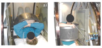
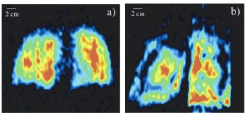
Walsworth and colleagues realised that patients who have inhaled spin-polarized gases do not need to be studied with the large magnetic fields of traditional MRI systems. Their new scanner therefore uses much lower magnetic fields of less than 10 milliTesla, which can be arranged around the patient in an ‘open geometry’ (see figure 1). This means that the patient does not have to lie down but can be studied standing up or sitting down, which also allows the lung to be studied in more detail (see figure 2). Patients have to breathe in hyperpolarized helium-3 gas through a plastic tube and a typical scan lasts just 30 seconds.
The team is now building a second-generation system with more homogeneous magnetic fields, which they will use for research on pulmonary physiology and respiratory diseases. “Eventually, portable low-field MRI scanners may be possible,” Walsworth told PhysicsWeb. “These could image the lungs of patients too ill to visit a conventional MRI machine – like premature babies, who often suffer from lung ailments.” The system could also prove useful for patients with pacemakers, who cannot be exposed to the high fields of conventional MRI scanners.
Meanwhile, another US group, led by Alex Pines at the University of California at Berkeley, has discovered a new technique – called remote detection – that has significantly improved the image resolution and sensitivity of MRI (J A Seeley et al. 2004 Journal of Magnetic Resonance 167 282). The technique depends on physically separating the two basic steps of magnetic resonance – signal encoding and detection. The scientists used laser-polarized xenon gas as the medium for ‘memorizing’ the encoded information, which was then carried to a remote detection site. The team improved the image resolution of MRI by several orders of magnitude and also increased the overall sensitivity of the technique.


