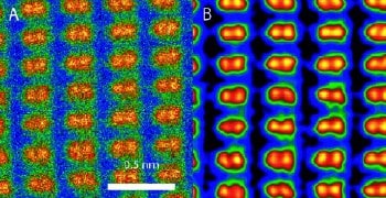
Scientists have imaged a crystal on sub-Angstrom scales by exploiting a new technique to correct the aberrations in a scanning transmission electron microscope. Although microscopists realized 50 years ago that it would be possible to make these corrections, the technology needed to do this has only just been developed by researchers at the Oak Ridge National Laboratory in Tennessee and Nion, a company based in Washington state (P D Nellist et al. 2004 Science 305 1741).

Sub-Angstrom imaging has been a long-standing goal for electron microscopists because it would allow structures to be studied at the level of single atoms. Although sub-Angstrom information can be obtained by post-processing electron micrographs, it has not been possible until now to obtain this information directly.
A scanning transmission electron microscope builds up an image by scanning an electron beam across a sample and measuring the parts of the beam that are transmitted back from the sample. However, the aberration — or blurring — that is caused by the magnetic lenses that are used to focus the electron beams has limited the resolution of these instruments to around 1.5 Angstroms (1.5×10⁻¹⁰ metres), which is slightly larger than the typical distance between atoms.
The resolution of an electron microscope increases as its aperture becomes larger but in the past lens aberrations have caused the images to blur once the aperture has reached a certain size. To overcome this problem, Stephen Pennycook of Oak Ridge and colleagues fitted an aberration corrector, made by Nion, onto their scanning transmission electron microscope. This corrector is a lens that employs software that is capable of analysing all axial aberrations in the microscope in less than a minute, and then making automatic adjustments to compensate.
The Oak Ridge-Nion team tested its device by imaging a silicon crystal in which the columns of atoms are known to be 0.78 Angstroms apart. Before correction, the optimum resolution for images was 1.3 Angstroms, but after correction it was possible to resolve the individual columns of atoms (see figure). The technique allowed the team to effectively double the aperture size of the lens.
“You can think of aberration correction as a pair of spectacles for a microscope,” says Pennycook. “Previously the vision was blurred but now we can see twice as clearly as before.”
“Seeing atoms more clearly allows us to see materials better, to understand how atoms go together so we can understand how things behave the way they do,” he adds. “The advance benefits a vast array of fields, from chemical sciences, material science and nanotechnology — everywhere people want to see what they have made.”
The team now plans to explore the possibility of using its device to image in 3D.


