Scientists in the US have demonstrated a new technique that is capable of starting crystallisation from scratch and then controlling and imaging the process as it proceeds in real time. Chad Mirkin and colleagues at Northwestern University in Illinois used an atomic force microscope coated with a polymer to grow crystals of the polymer on a mica substrate (X Liu et al. 2005 Science 307 1763).

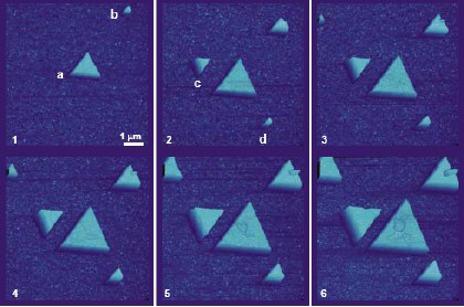
Mirkin’s team began by depositing a tiny drop of poly-DL-lysine hydrobromide (PLH) onto the mica substrate using the tip of an atomic force microscope (AFM) at room temperature. Next, they scanned the tip over the mica surface, covering an area measuring 8 microns by 8 microns, and observed that two triangular-shaped crystals, one of which was just 320 nanometres long, had formed. Then, as they continued to scan the tip across the surface, they saw these two “seed” crystals get bigger and other new crystals form (figure 1).
Overall they found that both the in-plane and out-of-plane growth rates of the crystals could be controlled by scanning the polymer-coated microscope tip across the surface. Control experiments, in which the mica was simply exposed to a solution containing the PLH, resulted in a haphazard formation of crystals with amorphous structures and triangles of various sizes.
When the temperature was increased to 35°C, the team observed that the triangular-shaped prisms changed into cubic-shaped ones (figure 2). Moreover, the size of the smallest crystal they studied was five orders of magnitude smaller than the minimum size that can be studied by X-ray diffraction techniques. This means that new features of crystallisation, which were previously too small to be detected, could now be observed for the first time.


