When the Dutch scientist Antonie van Leeuwenhoek first demonstrated the potential of the microscope in the 17th century, medicine was without vaccination, anaesthetic or antiseptic. Three centuries later, medicine has become far more specialized, but the microscope — crucial to many sub-disciplines like cell biology and pathology — has remained largely unchanged.
For physicians in the West this may not be a significant problem, but in developing countries diagnostic labs in the field struggle to equip themselves with conventional microscopes, which are both large and costly.
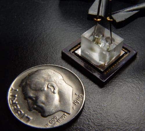
Now, scientists at the California Institute of Technology (Caltech) have developed a microscope the size of a penny piece that matches the resolution of its larger counterparts. What’s more, they claim it could be produced for as little as £5 a pop. Bioengineer and study leader Changhuei Yang told physicsworld.com that his new invention was inspired by the “floaters” in our eyes.
Direct projection
Floaters are small clumps of cells that have broken loose from the eye’s inner lining and drift through the watery gel known as the vitreous humour. We glimpse floaters as dark dots or scratches that occasionally enter into our field of vision.
Normally we see the world because light reflected from objects is focussed through the eye by a lens onto the retina. However, we see floaters by a different mechanism. These cells sit behind our lens and accumulate on the retina, and so we only obtain a scan or “direct projection” of them. Because the dots appear larger than the cells themselves, our body has a natural microscope that needs no lens.
In the Caltech replica of this effect, the specimen to be magnified is placed directly onto a complementary metal-oxide semiconductor (CMOS) sensor, which converts optical images into an electrical signal. Direct projection using this method was first demonstrated in 2005 by Dirk Lange of Stanford University, but so far resolution has not competed with conventional microscopes. At best the resolution has been the size of the pixels, or around three microns.
Yang and his colleagues get around this limitation by laying a thin film of aluminium over the sensor and then piercing it over the centre of each pixel. This simple idea restricts pixel sensitivity to the areas directly beneath the holes — effectively creating a smaller pixel that can match conventional microscope resolution. The researchers then suspend the specimen in an “optofluid” and let it flow into the holes.
‘Immediate application’
So what do the medics make of the pocket-sized microscope? “For diagnosis or screening of samples which contain macro parasites — such as worm eggs — this [microscope] has an almost immediate application,” says Dr Chris Drakely of the London School of Hygiene and Tropical Medicine. Unfortunately, Drakely warns, the microscope’s magnification factor of just 20 means it is not yet suitable for detecting malaria parasites, which requires magnification to be five times greater.
Yang recognises the need to further improve resolution but says his next plan is to pack several sensors into the same chip. If an array of thousands of microscopes could return data to the same interface, users could view a large sample but easily switch to a focussed view.
The tiny microscopes could also be inserted under a patient’s skin to monitor the spread of cancer. Some forms of cancer spread by tumour cells entering the blood stream, a process known as metastasis. Chips would be inserted in the appropriate region to “look out” for these cells. Yang points out, however, that this idea would be very difficult to implement, and at best the technology would be 10 to 15 years away.
Yang says that he has secured a patent and is “in talks with various multinational biotech companies”. He hopes his microscope will soon benefit developing countries, but does not want to take a major role in promoting the invention.


