An electron microscopy study by researchers in the US could provide important insights into the structure of shells, spines and other biological materials that are crystalline in nature. The team looked at how certain polymer strands are incorporated into a single crystal of calcite and found that the process involves physical interactions between the materials, rather than chemical reactions.
Single crystal structures are surprisingly common in living organisms. Each spine of a sea urchin, for example, is a single crystal of calcite. However, such spines have a very different shape than calcite crystals grown in the lab – and are often much tougher and more flexible. Understanding how these biomaterials are formed could lead to new types of manmade materials.
While scientists know that the properties of calcite are modified by the presence of organic molecules within the crystal lattice, it had proved difficult to obtain electron microscopic images of the interfaces between the calcite and organic molecules. Such images are crucial to understanding the nature of the composite materials. The problem is that electrons can only probe regions near to the surface of a sample, where the interfaces could be different than those in the bulk of a crystal.
Agarose hydrogel
Now Lara Estroff and colleagues at Cornell University have developed a new technique to study these interfaces. They began by growing a calcite crystal in a hydrogel containing agarose – a polymer that is found in seaweed and is known to incorporate into calcite.
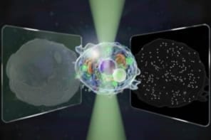
The team use an ion beam to carefully carve an extremely thin wedge-shaped pillar from the crystal – exposing the bulk of the crystal. The pillar is then placed in a transmission electron microscope (TEM) and a series of images is taken of a section of the pillar that is about 300 nm thick. The images are acquired at 2° intervals as the pillar is rotated through about 140°. Using computer tomography, the team is able to build up a 3D image of the crystal that shows exact locations of the agarose strands.
Electron diffraction data from the pillar confirmed that it was a single crystal – and allowed the team to determine the orientation of crystalline planes to individual agarose strands.
High-energy facets
The team found that the polymer strands sit next to “high-energy” crystal facets that would never be seen at the surface of a pure crystal.
Estroff told physicsworld.com that agarose is known to react very weakly with calcite, so it is unlikely that a chemical interaction is involved with this “stabilization” of high-energy facets. Instead she believes the stabilization is related to how a strand trapped in a growing crystal flexes and pushes back against its surroundings.
The team is now applying the technique to the study of biomaterials.
The research is described in Science.


