Germany’s Max Planck Society and the microscope maker Leica Microsystems have joined forces to commercialize a new technique to allow optical microscopes to look at even smaller objects than was previously possible. This latest method is based on stimulated emission depletion (STED) microscopy, which is a technique for beating the diffraction limit that was first demonstrated more than a decade ago by Max Planck researcher Stefan Hell.
Optical microscopy is the preferred tool of biologists because it can be used to study living cells in their natural environments. However, it does have one major limitation: it cannot normally resolve objects smaller than about 200 nm, the so-called diffraction limit. A major breakthrough came about a decade ago when Hell and colleagues at the Max Planck Institute for Biophysical Chemistry in Göttingen, Germany, invented STED.
The method involves fluorescence microscopy, whereby a molecule of interest is tagged with a dye that gives off light at a specific colour when illuminated with a spot of light. The sample is viewed through a confocal microscope, which uses a tiny aperture to ensure that light is only gathered from a very small part of the sample. The sample is moved under the aperture and many images are taken to build up a full picture.
De-exciting the dye
Normally, the resolution of the technique is given by minimum size of the spot of irradiating light – which can be no smaller than the diffraction limit. However, Hell came up with a clever way of narrowing the effective width of the light spot. This is achieved by firing a second beam of light that overlaps the spot. However, unlike the original spot, the intensity of this second beam is greatest at the outer edge and drops off rapidly towards the centre. The second beam de-excites the dye via stimulated emission, which prevents fluorescence from occurring where the second beam is most intense. Because of the second beam’s intensity profile, fluorescence only occurs at the centre of the light spot. This region, which resembles the hole at the centre of a doughnut, is much smaller than that allowed by the diffraction limit.
A STED module can be used with a conventional scanning confocal microscope to achieve a lateral resolution of about 50–70 nm – and a commercial version is available from the microscope manufacturer Leica.
Now, Hell and colleagues have found a way to improve STED to produce even sharper images. Normally, STED images are taken in a continuous wave (CW) mode. However, the researchers have discovered that it takes several nanoseconds until the STED doughnut has effectively depleted the previously excited molecules in the outer parts of the doughnut. This means that any images taken during this initial period are blurry because of the signal still coming from undepleted areas in the outer parts – and such images do not benefit from the full STED effect.
Rejecting early light
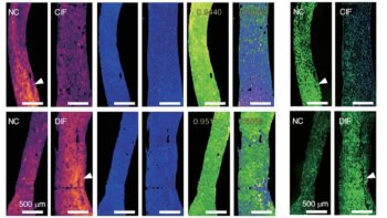
To get around this problem the team has invented gated STED – or g-STED – which rejects this early light. In doing so, the team was able to obtain much sharper images (see figure) and resolve structures smaller than 50 nm.
Another benefit of the technique is that sharper images can be acquired using much less light – and therefore less image-acquisition time. This means that it could be used to record the motions of molecules. Also, the fact that much less light can be used to obtain a sharp image means that it is less likely that living-cell targets would be damaged during irradiation.
Both of these capabilities will be welcomed by STED users, according to Paul O’Shea of the University of Nottingham in the UK. “There have been some worries that the photon dosages of imaging techniques such as STED can lead to cellular damage, so the g-STED approach is very welcome. Similarly, if the time and spatial resolution of the measurements with a gated acquisition allows the acquisition of FCS types of data, this would improve the information content of the imaging,” says O’Shea.
The commercialization agreement also involves the German Cancer Research Center. The new product will be launched in the first half of 2012 and Leica says that existing STED microscopes can be upgraded to perform g-STED.
The g-STED technique is described in Nature Methods 8 57.