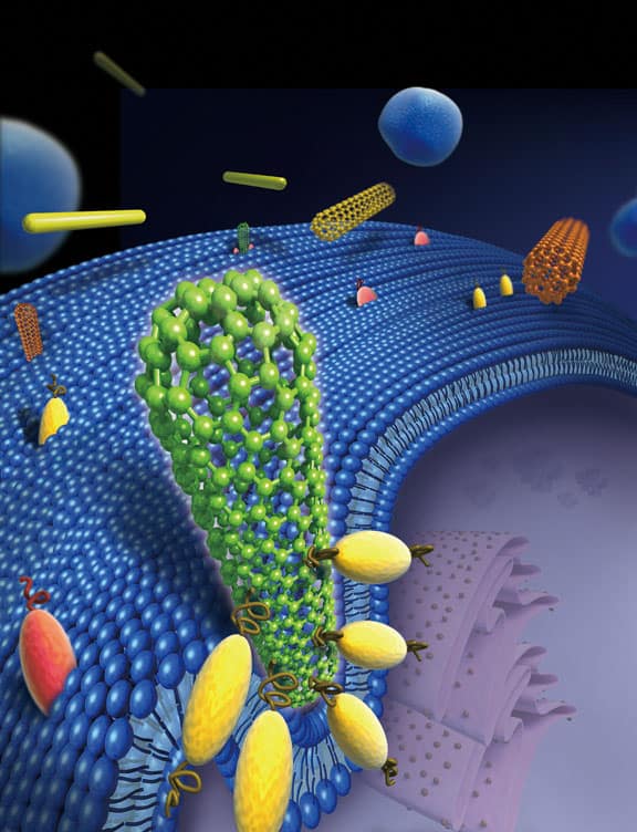
A new study by researchers in the US reveals that nanometre-thick strands such as nanotubes and nanowires enter biological cells head-on and almost always at a 90° angle. This orientation means that a cell mistakes the long cylinder for a sphere and tries to ingest it – with dire consequences. The findings could be important for designing safer, less-toxic nanomaterials.
Biological cells ingest objects in their environment by engulfing them, in a process known as endocytosis. The particles are taken into the cell, or “internalized”, and subsequently degraded.
However, things do not quite work out this way when it comes to very thin tubes, wires and fibres, say Huajian Gao and colleagues at Brown University. Optical-microscope techniques such as in situ “spinning disk confocal microscopy” have already shown that multiwalled carbon nanotubes enter cells head-on, or tip first. Now Gao’s team has now gone a step further by using ex situ field-emission scanning electron microscopy, which provides resolution on the nanoscale, to confirm these observations.
Similar to asbestos
The researchers have also found that most nanotubes enter a cell perpendicular to it. Indeed, a nanotube placed at a shallow angle (say 30°) to a cell membrane will actually rotate so that its tip ends up lying closer to 90°. Only then will the tip be engulfed by the cell. Such “tip entry”, as it has been dubbed, also occurs for other materials the team tested, such as gold nanowires and crocidolite asbestos fibres.
Gao and co-workers believe that the cells mistakenly treat the nanotubes as sphere-shaped particles rather than long cylinders. “But, by the time the cell realizes that the nanotubes are too long to be fully ingested, it is too late,” explains Gao. “Once the engulfing process begins, there is no turning back – within minutes, a cell senses that it cannot fully engulf the nanostructure and it calls for ‘help’; that is, it triggers an immune response that can cause inflammation.”
To test the theory, the team carried out so-called coarse-grained molecular dynamics (CGMD) simulations of how nanotube tips interact with a model lipid bilayer. The researchers found that receptors (which are made of proteins in real biological cells) diffuse along the bilayer and cluster around the nanotube. The receptors then adhere to the nanotube surface, pulling the tube into the cell as the bilayer wraps around the nanotube tip.
Minimizing energy
The likely reason for why the cell rotates the tube to 90° is that this angle seems to reduce the amount of energy needed by the cell to engulf the tube. According to the researchers, the rotation is caused by a torque exerted by the curved bilayer on the partially wrapped tube.
The researchers repeated their simulations for nanotubes with different diameters and lengths, and found that in the main the entry mechanism was the same. They did notice, however, that nanotubes with open ends – that is those with their caps cut off – did not enter cells at all but instead lay on the cell membrane. This is because open tubes lack the carbon atoms to bind to the receptors on a cell membrane, says Gao.
“Our work shows that tip geometry plays an important role in cell entry and can govern how cells and 1D nanomaterials interact with each other,” says Gao. “This leads us to pose the interesting question of whether modifying tube tips might help avoid such ‘frustrated’ uptake.”
Nanomaterials becoming ubiquitous
Understanding how nanomaterials interact with cells is crucial as nanotechnology becomes ever more present in our lives. Materials such as nanotubes, for instance, show great promise for next-generation microchips, composites, coatings, biosensors and, importantly, as drug-delivery vehicles.
Ken Donaldson of the University of Edinburgh in the UK, who was not involved in the work, says that more experiments are needed to confirm these results. “This study is interesting but is largely based on modelling – calculations on what nanofibres might do when they encounter a cell membrane. Would this be a mechanism that actually occurs in real life?” he asks.
Donaldson’s team has also studied nanotube–cell interactions and has obtained images that show some nanotubes entering a cell end-on but others that do so from the middle, and others still that do not enter at all.
Gao’s group published its results in Nature Nanotechnology 10.1038/nnano.2011.151.


