An endoscope made from a single optical fibre just 200 μm thick has been made by researchers in South Korea and the US. The device offers the possibility of imaging parts of the body where no endoscope has gone before; in addition, its powerful image-processing algorithms may also allow the device to take holographic images. While the method currently has several drawbacks – it will not work if the fibre is bent significantly, for example – it could find use in a number of medical-imaging applications.
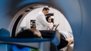
Endoscopy is a widely used medical procedure that involves a thin – and often flexible – tube being inserted into the body to obtain images of internal tissue. The fibre carries light into the body and then back out again. Although there are several different types of endoscope, they all need to have a way of illuminating the tissue of interest and a way of transmitting the image outside of the body. In situations involving very delicate tissues, making the endoscope as thin as possible could offer medical benefits.
In principle, an endoscope could be made from just a single optical fibre, so that a fibre with a 200 μm diameter, say, would be able to collect and transmit an image that covers a 200 μm-diameter circle on the subject. The problem with using just a single fibre, however, is that some of the light travelling down the fibre would be scattered by defects and get distorted beyond recognition. But what Wonshik Choi and colleagues at Korea University in Seoul have now done – together with researchers at the University of Pennsylvania and the Massachusetts Institute of Technology – is to find a way of characterizing these scattering processes and using this information to reconstruct the image.
Keeping track of scattering
Before a single fibre is used as an endoscope it has to be calibrated outside the body by firing a laser into one end of the fibre and measuring the brightness and phase of the light that emerges at the other end using a special camera. This process is repeated over a range of incident angles, with the resulting data being used to create a “transmission matrix” that describes how light makes its way through the fibre.
Although this matrix is enough to reconstruct an image that emerges from the fibre, the problem when the endoscope is used inside the body is that the tissue is illuminated by a laser beam sent down the fibre. This light also scatters, meaning that the tissue is illuminated with a “speckle” pattern of light and dark patches. So to ensure that the tissue is fully illuminated, the incident angle of this laser also has to be varied – effectively scanning the speckle pattern across the tissue.
Holographic bonus
One bonus of obtaining multiple data over a range of incident angles is that the information can be used to build up a holographic image of the tissue without having to scan the tip of the fibre. The team tested the method by showing that it can resolve a standard test pattern that contains a series of shapes, some as small is 2.2 μm across. The researchers then used the set-up to image the intestine tissue of rats, where they were able to use holography to study 3D structures in the tissue.
Low-cost endoscopes could be life-savers in developing countries
Allard Mosk, University of Twente
One important drawback of the endoscope – according to Choi – is that the fibre cannot be bent significantly from the shape at which the calibration was performed. As a result, the endoscope must be rigid – and not able to take full advantage of the fibre’s flexibility. Choi told physicsworld.com that the team is therefore exploring several solutions.
“The most promising approach, in my opinion, is the in situ calibration of the fibre,” he says. This involves injecting the fibre into the tissue while allowing it to bend. The transmission matrix for that specific curvature would then be obtained by firing a laser beam down the fibre measuring the light that reflects back up the fibre from the inner surface of the fibre tip.
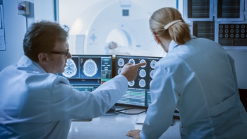
Diagnosing disease
The team is also developing a rigid endoscope and Choi says that in addition to its extreme thinness, the device can obtain images with higher spatial resolution than existing instruments. “We can attain a resolution below 1 μm and this this will facilitate in vivo disease diagnosis,” he says.
Allard Mosk of the University of Twente in the Netherlands believes that an important advantage of Choi’s endoscope is that it combines a simple design with computer processing. “Using a computer to correct for the optical distortions is very much cheaper when mass produced than making high-quality optical endoscopes,” he explains. “Low-cost endoscopes could be life-savers in developing countries,” he adds.
Mosk and colleagues have recently developed a way of obtaining images through biological materials that are normally opaque to light. Their technique also involves scanning speckles across an object and Mosk suggests that it could be combined with the endoscope to allow for an even clearer view – particularly when the tip cannot be brought right up to the tissue of interest and the light passes through intervening material.
The endoscope is described in Physical Review Letters.