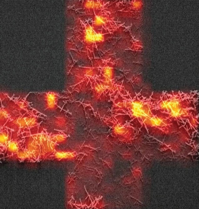
Tiny “tags” made of dye molecules stuffed into carbon nanotubes have been used to develop a high-resolution imaging technique based on Raman scattering. Created by researchers in Canada, the tags boost the weak Raman signal of molecules about one million times. The new approach could lead to improved medical diagnostics and treatments, and could even be used to fight counterfeiting.
Raman spectroscopy involves shining a beam of light onto a solid or a liquid to identify its molecular composition. While most of the photons will scatter from the sample with no change of energy, a small number of photons will exchange a tiny amount of energy by causing molecules in the sample to vibrate. This is called Raman scattering, after the Indian physicist Chandrasekhara Venkata Raman who first observed it in liquids in 1928 and won the 1930 Nobel Prize for Physics for his discovery.
“One can imagine Raman scattering as the process by which photons shake a molecule,” explains Thomas Szkopek of McGill University, who is part of the research team. The vibration of each type of molecule is unique and so is the amount of energy exchanged with photons. By measuring the energy spectrum of the scattered photons, it is possible to determine what molecules are present in the sample.
Major limitation
Raman spectroscopy is widely used in medicine, chemistry and drug development. A major limitation, however, is that the scattered light is very weak. This makes the technique impractical for many applications, especially those involving high-resolution imaging of biological samples, because it takes so much time to collect the weak signal from each pixel.
Increasing the strength of the interaction between the light and the molecules does not help much, as it leads to the Raman light being hidden by much brighter fluorescence. “To be practical in high-resolution imaging, there is a need to boost the Raman signal by about a million times or more,” says Richard Martel of the University of Montreal, who led the research team.
There have been previous attempts at amplifying the Raman effect, but most of these involved manipulating the optical field with metal nanostructures. This requires putting the molecules of interest in direct contact with metallic particles, but this has proved difficult because the particles are difficult to stabilize and control.
We can turn up the brightness of the Raman light while leaving the fluorescence light turned off
Thomas Szkopek McGill University
Martel’s team, which includes postdocs Étienne Gaufrès and Nathalie Tang of the University of Montreal, took a different approach and turned to dyes and carbon nanotubes, which are hollow tubes of carbon with walls as thin as just one atom. “The carbon nanotube is very effective at turning off the undesired dye-molecule fluorescence while allowing the desired Raman scattering process to continue,” explains Szkopek. “We can turn up the brightness of the Raman light while leaving the fluorescence light turned off.”
Tubes filled with dye
The researchers begin by using nitric acid to purify the nanotubes, cleaning them and opening their ends. They then dissolve dye molecules in a solvent, mix in the nanotubes and heat the solution for a few hours. The dye molecules fill the tiny nanotubes and align along the tube axis. The resulting “nanoprobes” are about 1 nm in diameter, 500 nm long and contain about 500 dye molecules.
The next step involves adding a nanoprobe to the sample to be examined. The nanoprobes can be chemically grafted onto any object, even bacteria or proteins, therefore becoming a sort of “Raman tag”, says Martel. “Attaching the nanoprobes to a target is like printing a barcode into the object, allowing it to be identified even if it’s not Raman active or visible,” he says.
A measurement then proceeds like a normal Raman spectroscopy procedure. A laser beam inserted in the microscope objective shines onto the sample and its scattering is probed with a spectrometer. The only difference is a huge increase in the intensity of the Raman signal.
Protected from photobleaching
“Once the dye molecule absorbs a photon, the energy is passed on to the nanotube before the molecule has the opportunity to change the energy back into a photon that would contribute to fluorescence,” says Szkopek. Carbon nanotubes therefore suppress the fluorescence that would otherwise overwhelm and hide the Raman signal, making the signal from the nanoprobes so bright that it completely dominates the spectrum – one million times stronger than the Raman signal of other molecules, once the fluorescence is supressed. The nanotubes also protect the dye from the environment, especially photobleaching by the laser beam. This tagging approach is already used in fluorescence, Szkopek adds, but the fluorescence signal is not a great barcode compared with the Raman signal – many more complex signals can be generated with the dyes in a Raman nanoprobe.
There are other advantages to using the technique, says Mark Hersam of Northwestern University, who was not involved in the study. For instance, the ability of the nanotubes to supress fluorescence and at the same time to protect the dyes from the environment allows for broad use of such Raman tags – in particular, “for multispectral analysis in applications ranging from protein detection to biomedical imaging”, Hersam says. He also thinks that nanoprobes could be added to banknote ink could help eliminate the problem of counterfeiting.
The research is described in Nature Photonics.


