A new detector that could improve the effectiveness of proton-beam cancer therapies has been developed by researchers in the UK and South Africa. The system tracks protons that have travelled through the body and gives medical physicists a detailed picture of how a therapeutic proton beam will interact with the treatment area. Having this information could lead to better cancer therapy and the team plans to build a prototype scanner that could eventually be commercialized.
Protons are ideal for some cancer treatments because when fired into living tissue, a beam of protons deposits most of its energy at a very specific depth that depends on its initial energy. As a result, protons can be used to destroy tumours while leaving surrounding healthy tissue relatively unharmed.
Before a patient can receive treatment, medical physicists must calculate the appropriate radiation dose distribution to be delivered by the proton beam. Normally this is done by doing a conventional X-ray computed-tomography (CT) scan of the treatment area and using this information to calculate how much energy will be absorbed from the proton beam – a quantity called the “stopping power”. However, uncertainties can arise in these calculations and therefore medical physicists are keen on developing better methods for determining the stopping power.
Researchers at the Proton Radiotherapy Verification and Dosimetry Applications (PRaVDA) consortium – funded by the Wellcome Trust – are designing and building the first proton transmission CT scanner based on silicon-based CMOS active pixel sensor (APS) technology. Such scanners work by exposing the treatment area to a beam of protons and then detecting the protons that have passed through the body. This information is used to build up a 3D image of the region to be treated, which provides an accurate measure of the stopping power. The benefit of using these APS detectors over existing calorimeter detectors is that they can track more than one proton at a time, which reduces the amount of time needed to perform a scan.
Localized interactions
In their latest work, the researchers have shown that their DynAMITe sensor can resolve individual protons passing through it. Developed by PRaVDA researchers in a previous project, the radiation-hard pixellated sensor has a 12.8 × 12.8 cm area and two wafer diode layers, one with 100 µm pixels and another with 50 µm pixels. The pixelated design allows proton-sensor interactions to be localized within the sensor area.
“This allows you to measure the passage of more than one proton in the device at once,” explains team member Gavin Poludniowski, a medical physicist at the University of Surrey. The capability is an advantage over calorimeter-based sensors that handle one proton at a time. “[For these] it has been a challenge to get the event rate high enough to take a scan in a practicable time,” explained Poludniowski.
Telescopic tracking
In the planned proton CT scanner, a stack of the CMOS sensors – essentially a “telescope” – will determine proton energy loss in the patient. Combined with other detectors that were not a subject of this study, data from the telescope will also determine the direction of protons exiting the patient. With this information, the path of the proton in the patient – that is a result of multiple coulomb scattering events – can be reconstructed, generating images with superior spatial resolution to those achievable by detectors that assume an unscattered, linear trajectory.
The researchers demonstrated the sensor’s proton counting ability by irradiating it with a 36 MeV beam produced by the MC40 cyclotron at the University of Birmingham in the UK and a therapeutic 200 Mev beam at the iThemba treatment facility in Somerset West in South Africa. Low beam currents and high frame rates of 1400 Hz – achieved by reading 10 of the 2520 rows on the sensor – maximized the ability of the sensor to resolve individual proton interactions.
Detected events increased linearly with beam current up to a nominal current of 0.1 nA, then fell off with further increases. The observation is consistent with pulse pile-up in the sensor pixels that, in turn, indicates the detection of individual protons. Experimental observations also agreed with Monte Carlo simulations of the same set-up, providing further evidence of proton counting by the sensor.
Stacks of DynAMITe
When two DynAMITe sensors were stacked together – double DynAMITe – event distributions in the two matched. Eliminating fluctuations in beam current as a confounding factor, the high correlation that the researchers observed (r = 0.854) indicated that the pair was detecting the same protons, confirming its tracking ability.
With proof-of-concept established, the researchers are redesigning the DynAMITe sensors for improved performance, with increased frame rates a major focus of their efforts. Estimating that the proton CT scans will require tens of millions of image frames, their goal is to achieve a 1000 Hz frame rate for the readout of the entire sensor area to limit scan duration to a few minutes.
“We are investigating various aspects of hardware design to get the frame rate that we need. Pixel size and bit-depth are factors,” says Poludniowski. “Substantial innovations are [also] being made in the read-out design and electronics.”
First scans in late 2015
Investigations into the effects of telescope geometry on performance and the radiation hardness of the sensor are also in progress. The consortium plans to build a device and perform the first scans by the end of 2015, and then commercialize the technology with an industrial partner.
The research is described in Physics in Medicine and Biology.
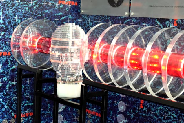
- PRaVDA researchers are exhibiting their work at the Royal Society’s Summer Exhibition in London from the 1–6 July.
- This article first appeared on medicalphysicsweb.org.


