Researchers in the US have used a self-assembly method based on viruses and DNA to position almost 200 fluorescent molecules to within a few nanometres of a tiny gold nanoparticle. This accurate positioning of the molecules boosts their fluorescence output and the method could have applications in information processing, sensing and energy technologies.
Electrons in a metallic nanoparticle undergo collective oscillations known as a surface plasmon resonance when exposed to certain frequencies of light. The nanoparticle then behaves as a tiny antenna, concentrating light within a few nanometres of the nanoparticle surface. If a fluorescent molecule – or fluorophore – is placed within this region, the amount of light captured by the molecule can be boosted significantly.
“This provides a means of creating an intense electromagnetic field near a light-absorbing centre, thus allowing us to greatly increase the capture of light,” explains James De Yoreo of the Pacific Northwest National Laboratory, who was part of the research team. Boosting the number of fluorophores surrounding the nanoparticle helps increase the amount of light captured even further. However, the process is very sensitive to the distance between the nanoparticles and fluorophores, making it difficult to take advantage of the effect in complicated arrangements of nanoparticles and fluorophores.
Nanoscale assembly
The researchers, from the Lawrence Berkeley National Laboratory, the Pacific Northwest National Laboratory, University of California, Berkeley and Arizona State University, combined two self-assembly approaches to collect hundreds of fluorophores and position them next to a gold nanoparticle.
First, they used a virus “capsid” to form a container for the fluorophores. The capsid, which would normally encapsulate a virus, is made of a protein that self-assembles with many copies of itself to form a shell. The researchers modified the inner surface of the shell with fluorophore attachment sites, capturing almost 180 fluorophores and giving a density of one fluorophore per 14 nm2. The capsid was further modified so that the outside was coated with DNA strands.
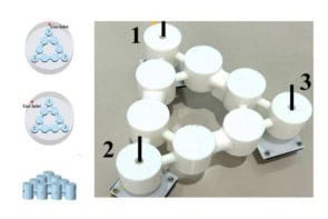
The second step involved a process called “DNA origami”, whereby a collection of several hundred synthetic DNA strands self-assemble into shapes that are around 100 nm in size. The researchers formed a tile to act as a “molecular breadboard”, with two binding locations to allow objects to be attached. Finally, the team modified gold nanoparticles with a set of DNA strands that recognized a location on the origami. The DNA sequences on the outside of the capsid were designed to bind to a different location.
Adjustable origami
Mixing the three components together – the fluorophore-loaded capsid, the origami tile and the gold nanoparticle – resulted in the capsid and nanoparticle being held nanometres apart on the origami. By adjusting the origami design, the capsid–nanoparticle separation distance could be adjusted.
The researchers confirmed that the system had formed as designed by using atomic force microscopy and electron microscopy. First they investigated how the capsid–nanoparticle separation affected the fluorescence characteristics by studying a sample using confocal microscopy. The separation distance was then determined using atomic force microscopy. The combined measurements demonstrated increased fluorescence intensity for a number of separation distances.
To better understand the experimental results, the researchers modelled the interactions of the fluorophores with the nanoparticle, demonstrating that the system behaved as they expected. For larger sizes of gold nanoparticle, the model showed that the fluorophores would undergo significant increases in fluorescence.
Mix and match
“The model enabled us to explore changes to the nanoparticle size, choice of fluorophore, arrangement of fluorophores and even the capsid shape to optimize the performance,” says De Yoreo.
The primary interest of the team is to create technologies that mimic some of the highly efficient processes that living organisms use to harvest energy from the Sun. “Our use of the effect is directed towards energy harvesting for solar-energy applications,” explains De Yoreo. “When one looks at light-harvesting complexes in biological systems, they often utilize a similar architecture.”
The research is described in ACS Nano.
- This article first appeared on nanotechweb.org


