A new laser-based scanning technique, which could potentially help researchers to get new information from fossil specimens, has been developed by scientists in the US. The inexpensive and non-destructive approach uses commercial-grade lasers to stimulate fluorescence in the fossil, revealing detail that would not otherwise have been observable with traditional visual enhancers such as UV light, which have a far lower irradiance level. In palaeontology, a variety of visual enhancers have long been used to highlight fossils for photography and analysis. One interesting technique uses UV light, which can stimulate visible fluorescence in certain minerals such as hydroxyapatite (the inorganic component of bone) – and, in some cases, may even highlight fossilized soft tissues. Most minerals in fossil specimens are hard to fluoresce, however, which means that, under UV light, they appear to remain dark.
Fluorescing fossils
The intensity of fluorescence can be increased, however, by using a higher-powered light source such as a laser, which allows for detectable fluorescence from a far wider variety of specimens. While laser stimulation has been traditionally constrained to highly detailed studies on microscopic scales – either through confocal laser scanning microscopy or Raman spectroscopy – recent developments and cost reductions in commercial laser technology have allowed palaeontologist Tom Kaye of the Burke Museum in Seattle, and colleagues, to apply laser-stimulated fluorescence to the macroscopic level.
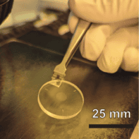
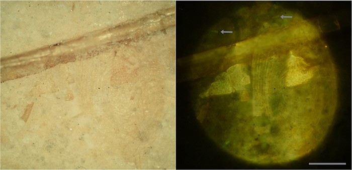
The method is quite simple – in a darkened room, fossil specimens are excited by laser light and viewed through an appropriate long-pass filter. The filter blocks the bright laser light but allows the fluorescent signal from the fossil specimens to pass through. This can then be photographed as a long exposure with a digital camera. Different wavelengths excite different rocks and fossils in different ways. Indeed, even if a particular fossil will not fluoresce, it may still be possible to illuminate the surrounding rock and backlight the specimen.
Rocky specimens
“We are excited about this technique because it offers instantaneous geochemical fingerprinting of the specimens,” says Kaye, explaining that the technique will enable the researchers to identify a variety of soft tissues preserved in fossils, adding that they will be able to “look at things like skin, the size of muscles and the construction of feet”.
Alongside revealing additional detail in existing specimens, the laser light is also powerful enough to penetrate a limited distance into certain rocks, allowing the team to visualize fossil specimens that may be entirely or partly hidden beneath the rock’s surface. Using their laser technique, the researchers were able to easily identify a 120 million-year-old, largely encased “mystery fossil” – a fish – when the specimen’s teeth and bones fluoresced at a higher intensity than the surrounding rock matrix.
Fossil fakers?
Laser scanning can also help identify composite fakes – fossils that have been cobbled together from different specimens – by revealing differences in fossil mineralogy. “People try to make the specimens look better or more intact because this makes them easier to sell,” explains team-member David Burnham of the University of Kansas. “Some artists are so good that you can’t tell where the real thing stops and the fake thing begins. With lasers, now we’ll know.”
The researchers have even applied the fluorescence-based technique to create the world’s first automated fossil sorter. Rock grains are sent, in a narrow stream, past a laser and video camera; based on their fluorescence, potential small fossils (for example teeth) are separated for closer examination. The machine can process 1–2 kg of material per hour – an amount that would take weeks or months by sort hand – producing a specimen concentrate of typically 20–50%.
“Laser-stimulated fluorescence is an important, welcome addition to the now rapidly expanding arsenal of techniques available for modern studies of ancient life,” says William Schopf, a palaeobiologist at the University of California, Los Angeles, who was not involved in this study. He told physicsworld.com that although the method is incapable of producing “the high-resolution 3D cellular detail provided by confocal laser scanning microscopy, and does not provide the sub-micron macromolecular information afforded by Raman spectroscopy, it is faster, cheaper, more easily portable and offers an order of magnitude improvement in the signal-to-noise ratio over traditionally used standard UV light”.
“It is splendid that both structural and anatomical detail can be rapidly resolved using this de novo approach,” agrees Phillip Manning , a palaeontologist from the University of Manchester in the UK. “I have no doubt that the imaging of fossils using multiple facets of the electromagnetic spectrum will shed unique light on the evolution of life on Earth.”
Having demonstrated the potential of laser-stimulated fluorescence, Kaye and his colleagues have moved to applying the tool to reveal new details about previously examined fossil specimens. The team is also exploring how the method could be used to scan an area for fossils, at a range of around 100 metres, by coupling a laser with a telephoto camera.
The research is published in PLOS ONE.