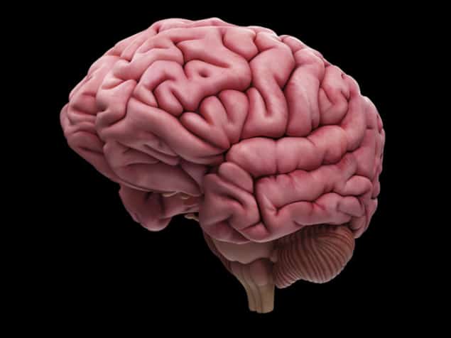
The distinctive folds of the human brain are the result of mechanical compression caused by growth during development, according to an international team of scientists. Using a 3D-printed gel model of the brain, the researchers have now shown that forces generated during expansion can create the brain’s wrinkled shape. This mechanical model was first proposed in 1975, but it has been difficult to test.
Highly folded brains are only seen in a small number of species, including some primates, dolphins, elephants and pigs. From an evolutionary perspective, the reason why the brain folds is quite simple – it maximises the number of neurons that can be squeezed into the space, while reducing the distance between them, which improves cognitive function. In humans, the outer layer of brain tissue – the grey matter or the “cerebral cortex” – starts to fold when the foetus is around 23 weeks old. The process where the cerebral cortex forms its folds, known as “gyrification”, continues until adulthood, when our brains stop growing. By this point the brain has increased about 20-fold in volume and 30-fold in surface area.
Grey-matter origami
While the purpose of gyrification is understood, the mechanism behind it is not. A number of biochemical and mechanical theories have been previously proposed, including one which suggests that folding is caused by mechanical tension generated in the neurons, but none have been proven. Tuomas Tallinen from the University of Jyvaskyla, Finland, together with colleagues in France and the US, now say the most likely explanation is the simplest one: the cerebral cortex expands faster than the rest of the brain, while changing little in thickness. Essentially, the cerebral cortex remains the same as its surface area grows. This produces compressive stress, which in turn leads to the mechanical folding of the cortex.
To test its theory, the team created a 3D cast of an unfolded 22 week-old human brain, based on MRI scans. This was used to create a gel model of the core of the brain, which was then coated in a thin layer of absorbent elastomer gel to represent the cerebral cortex. When immersed in solvent, the outer layer of the gel-brain swelled relative to the inner core and as the compressive forces built up it began to crumple. Tallinen told physicsworld.com that they “observed folding patterns that are qualitatively very similar to the folding patterns in foetal brains during the early stages of gyrification”.
After it had finished expanding, the model resembled a 34 week-old foetal brain. “The key parameters setting the size and qualitative appearance of the folds are the stiffness of the grey-matter top layer relative to white-matter substrate, and the thickness of the grey-matter layer relative to brain size,” says Tallinen. “More subtle is how the folds get oriented – the geometry of the foetal brain surface is a determinant for dominant orientations of the folds.”
Model material
Christopher Kroenke from the Oregon Health & Science University in the US, who was not involved in the research, says that similarities between the folds suggest that the “mechanical features of the model bear strong resemblance to those of the human brain” and that the “agreement between finite element calculations and experimental observations strongly supports that mechanical compression is the driving force behind folding in the model”. But he adds that further examination of the similarities and dissimilarities between the model material and foetal brain tissue “will be valuable for determining whether mechanical compression indeed drives folding of the cerebral cortex”.
David Van Essen from Washington University in St Louis, who first proposed the neuron tension-based theory, says that while the research “uses a clever combination of physical modelling and finite-element simulations to show that several features of human cortical folding can be emulated by a ‘buckling’ model”, it has a key limitation. “Their assumption that cortical thickness is fixed during massive tangential expansion is biologically implausible in the absence of a mechanism that could account for this highly anisotropic growth,” he explains. He adds that limited cortical thickness can be explained by mechanical tension along the neurons, as well as various patterns seen in the folds of the brain.
The research is published in Nature Physics.


