A device that can perform 3D ultrasound imaging with just one large piezoelectric ultrasound transmitter and receiver has been developed by researchers in the Netherlands. Most ultrasound imagers use thousands of tiny sensors to transmit ultrasonic bursts and record the reflected signal, but the new device uses an aperture coding mask in front of a single sensor instead. According to the scientists, this could pave the way for cheaper, faster, simpler and smaller ultrasound devices with new clinical applications.
Ultrasound is widely used in medicine to obtain rapid, real-time images of internal body structures. As well as monitoring development in pregnancy, it is also used to investigate health problems such as heart disease, soft-tissue damage and abdominal pain. Ultrasound is popular because it is low-cost compared to other imaging techniques and doesn’t use ionizing radiation. The imaging technique also has a wide range of industrial applications such as looking for cracks and other defects in materials.
Almost all ultrasound systems use arrays of large numbers of transducers that produce short ultrasonic bursts. In medical applications the bursts are reflected back by tissues in the body and detected by the same array. The time delay between transmission and detection and the strength of the reflected signal provide information on the location and density of the tissue, which is used to construct the image.
Extra dimension
Most ultrasound systems produce 2D cross-sectional images, but 3D images would be better for medical assessments. The problem is producing 3D images from current ultrasound techniques is not that simple. Most systems use transducers distributed along a single direction. To obtain a 3D picture the ultrasound needs to be moved around the tissue being imaged, which often isn’t possible. A more complicated solution is a 2D transducer array that steers the beam, which is an expensive piece of technology.
To tackle this issue, Pieter Kruizinga, a biomedical engineer at the Erasmus Medical Center in Rotterdam, and his colleagues, turned to compressive signalling, a processing technique that allows a signal to be decoded with less measurements than traditionally thought necessary. In a new paper in Science Advances, they describe how this has enabled them to produce an ultrasound device that can create 3D images using a single ultrasound transmitter and sensor.
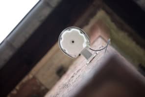
Rather than using an array to produce a focused, steerable ultrasound beam, their device uses a plastic mask that scrambles the ultrasound signal to produce a large scattered ultrasound field. “With this mask, we create such a chaotic signal that every point in space has its own signal attributed to it,” says Kruizinga. If you do this well enough, you can then separate and identify these signals when they return to the sensor, Kruizinga explains. These data – along with information on time-delay and signal strength – are then combined with detailed knowledge of the direction in which each unique signal was sent, and thus where it bounced back from, to build the image.
Unique pixels
The mask enables the creation of a single compressed ultrasound measurement of an object in which every single pixel in the image is uniquely identifiable and can be decoded using compressive signalling techniques.
“Without this mask you would have a very nice beam propagating through your medium, but then all pixels, every point in space, would receive the same signal,” Kruizinga says. “If you then receive back the signal you wouldn’t be able to know which points in space were active.”
To improve the image further, the researchers also rotated the mask in front of the sensor. This rotates the ultrasound pattern, allowing additional measurements and information to be obtained.
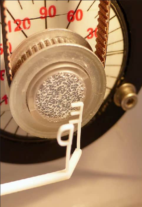
The team showed that the device can be used to 3D image two plastic 3D-printed letters (see figure). They say, however, that the device “does not deliver similar functionality as existing 3D ultrasound arrays”. But claim that the introduction of other techniques to introduce signal variation and improved predictions of the ultrasound field produced by the mask could lead to better results.
Easy life
Kruizanga told Physics World that they have already started testing devices with multiple transmitters. “We wanted to show that it is possible with one, but you can make more unique signals and life becomes a lot easier if you use a couple of them,” he explains.
Since publishing the paper, Kruizinga says that one of his colleagues has managed to shrink the device into a probe around 1 mm thick. He says that such a device could be used for intra-vascular imaging – to examine artery blockages and help with the placement of stents, for example.
As well as being easier to shrink for imaging inside the body than multiple transducer arrays, Kruizinga says that devices based around their technique will also be cheaper and more portable than current medical ultrasound machines.