Flash Physics is our daily pick of the latest need-to-know developments from the global physics community selected by Physics World‘s team of editors and reporters

Mapping Earth’s magnetism
The magnetic field of the Earth’s crust has been revealed like never before in a new high-resolution map. To produce the plot, Nils Olsen from the Technical University of Denmark (DTU) and colleagues have used a new modelling technique to combine data from the European Space Agency’s (ESA) Swarm satellites and their predecessor, the German CHAMP satellite. Presented at the ESA’s Fourth Swarm Science Meeting in Canada, the map is the highest-resolution plot to date of the crust’s magnetic field. While the majority of the Earth’s magnetic field is generated by molten iron in the core, a small part of it is created by magnetized rocks in the lithosphere (the crust and upper mantle). This “lithospheric magnetic field” is very weak and difficult to detect. Taking different orbits around the Earth, Swarm’s three identical satellites use “new-generation” magnetometers to measure the direction and magnitude of the field. The new map shows field features down to about 250 km, therefore highlighting anomalies such as one in the Central African Republic. The exact cause of this strong localized magnetic field is unknown, but Olsen and team speculate that it is the result of a meteor impact 540 million years ago.
Vibronic spectroscopy done at atomic resolution
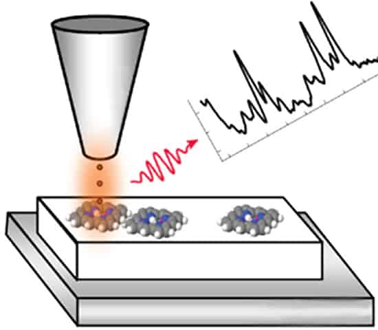
A new technique that combines the chemical sensitivity of optical vibronic spectroscopy with the atomic resolution of scanning tunnelling microscopy (STM) has been developed by Guillaume Schull and colleagues at the University of Strasbourg in France. Near-infrared, Raman and low-temperature spectroscopies involve the vibrational modes of molecules and can therefore provide important information about the chemical, structural and environmental properties of organic molecules. But because these are optical techniques, their spatial resolutions are normally limited to approximately the wavelength of the light used to probe the molecules – typically hundreds of nanometres. STM involves bringing an atomically sharp tip near to a molecule on a surface and measuring the electrical current that flows between the two. While STM can create sub-nanometre-resolution images of molecules showing individual atoms, it does not provide much chemical information about its subjects. Now, Schull’s team has come up with a way to do vibronic spectroscopy without illuminating the sample with light. Instead, electrons from the STM tip excite vibrational modes of molecules of interest, causing them to emit light that can then be detected. The team used the technique to study individual phthalocyanine molecules deposited on a surface and measured an intense emission of red light at 1.9 eV as well as several weaker signals at lower energies. Writing in Physical Review Letters, Schull and colleagues say that a comparison to data acquired using conventional Raman spectroscopy confirms that the STM-acquired spectrum provides “a detailed chemical fingerprint” of the phthalocyanine molecules.
Electrical “nerve cuff” could help treat chronic disease

A tiny device that is implanted in the body and delivers electrical signals to the nervous system has been created by Timothy Gardner and colleagues at Boston University in the US. Described as a “nerve cuff” or a “nanoclip”, the device is designed to stimulate nerves as part of the treatment of a range of disease including diabetes, polycystic ovarian syndrome, asthma and cancer. The implant is currently being tested in small animals and is therefore very small – measuring just 200 nm to target nerves that are as small as 50 μm in diameter. The devices are made using a laser writing technique, which the team says allows the nanoclips to be designed for use in keyhole surgery. The devices were tested by implanting them on the hypoglossal nerves of zebra finches. This is the nerve that control’s the tongue of the birds and it therefore plays a crucial role in how they sing. Writing in the Journal of Neural Engineering, Gardner’s team says that studies of the finch’s birdsong revealed no change due to the presence of the nanoclips. This, they say, suggests that the implant is safe to use.
- You can find all our daily Flash Physics posts in the website’s news section, as well as on Twitter and Facebook using #FlashPhysics.


