Fundamental breakthroughs in physics are continuing to yield new medical technologies for identifying and treating a range of diseases.
Throughout this century advances in physics and medicine have gone hand in hand. The most fundamental discoveries in physics have rapidly been exploited by the medical community to devise new techniques for diagnosing and treating a variety of illnesses. And physicists are increasingly listening to the demands of the medical profession when defining the direction of new research.
The best known example of the link between physics and medicine is the use of X-rays to diagnose and treat disease. Less than a year after their discovery by Wilhelm Röntgen in 1895 scientists had found that X-rays could help to treat malignant tumours and cancer. But the impact of X-rays was not fully revealed until the early 1970s, when an imaging technique known as X-ray computed tomography was introduced. This method – now known simply as CT scanning – made it possible to construct three-dimensional images of the human body for the first time.
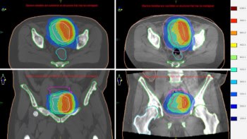
Recent research efforts have focused on improving the effectiveness of treating cancer with X-rays. As Steve Webb reports (see summary), malignant tumours can be destroyed by focusing several X-ray beams onto the diseased tissue. Progress in generating “shaped” beams has made it possible to match the high radiation dose to the shape of the tumour, but existing techniques cannot cope with the 30% of tumours that have dips or concavities in their surface. To solve this problem techniques are now being developed to vary the intensity of the X-ray beams by moving small pieces of lead or tungsten into the radiation field for precisely controlled periods of time. Such techniques are still at the research stage, and clinical trials are just beginning.
Hyperpolarization reveals all
Basic physics research has also played a crucial role in the development of magnetic resonance imaging (MRI). This technique is based on nuclear magnetic resonance, a quantum phenomenon that was first demonstrated by Edward Purcell and Felix Bloch in the late 1940s. Imaging systems based on this effect were first demonstrated in the 1970s, and MRI has since developed into a powerful clinical tool.
But even the latest equipment cannot image inside the lung. Conventional MRI detects the signals generated by the nuclear spins of protons present in water and fat, but the proton density of the lung is too low to generate clear images. In their article (see summary), Allan Johnson and colleagues explain how this problem can be overcome through the use of “hyperpolarized” gases, an idea that has emerged from experiments in atomic physics carried out over the last 30 years or so. Hyperpolarized samples of helium and xenon can be created with powerful lasers that induce atomic transitions in the gas and hence align some of the nuclear spins. This strengthens the magnetic resonance signal, making it possible to image the air spaces throughout the lung and to diagnose bronchial diseases such as emphysema. The technique is now being extended to monitor blood flow to the lungs and brain.
Nonlinearity and the heart
A less well known application of physics in medicine is the use of nonlinear waves to explain the motions of the heart. The cardiac muscle is essentially a mechanical pump that drives blood throughout the body. Electrical impulses sent from the brain propagate through the muscle, exciting the tissue and stimulating a synchronous contraction of the heart. This electrical excitation of the heart can be described in terms of a nonlinear wave equation that can be combined with biophysical models to simulate wave propagation in the heart.
These numerical studies are helping to understand what happens in a cardiac arrhythmia, a potentially lethal condition in which different parts of the muscle contract at different times. As Arun Holden explains, arrhythmias can be described in terms of “re-entrant” wave propagation, where the same wave of electrical activity repeatedly passes through the same tissue. Numerical models of the heart have revealed how these re-entrant waves can lead to a highly irregular motion of the heart known as fibrillation.
If fibrillation occurs, the traditional approach is to “restart” the heart with a single electric shock. However, the physical basis of this approach is not clear and the strength of the shock can damage the heart tissue. Studies of nonlinear wave propagation indicate that it may be more effective to apply a series of small-amplitude shocks. Another approach might be to modify the properties of the re-entrant wave with drugs.
These are just a few of the ways in which physics has been exploited in medicine. Optical-based techniques are widely used for imaging and analysis, while lasers are increasingly being employed for microsurgery. Ultrasound has also become a common clinical tool, and researchers are now attempting to exploit high-intensity ultrasound for bloodless surgery. Over 100 years since Röntgen discovered the X-ray, the collaboration between physics and the medical community continues to yield new treatments that improve the life of everyone.


