When James Watson and Francis Crick published the structure of DNA 50 years ago next month, it revealed the leading role played by the Cavendish Laboratory in structural biology. But why was this discovery made in a physics lab in Britain?

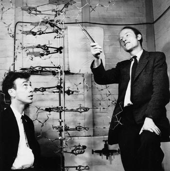
ON 25 April 1953 James Watson and Francis Crick, working in a small Medical Research Council unit in the Cavendish Laboratory, Cambridge, published a short letter in Nature. It described a remarkable two-chain helical structure for DNA, the biological polymer that constitutes the hereditary material in living organisms. The structural details of their model immediately suggested a mechanism by which the genetic material would replicate itself. Furthermore, the model indicated clearly what principle is used by the genetic material to store all the information needed to synthesize the proteins required to build a living organism.
In constructing their model, Watson and Crick were helped by advance knowledge of X-ray data obtained by Rosalind Franklin and Maurice Wilkins at King’s College London. These results – and additional data and reasoning supporting the Cambridge structure – were published in the same issue of Nature. The double-helix model provided the key to a detailed understanding of how living cells can produce two exact copies of themselves. It was a major factor in the enormous revolution in biology that dominated science in the second half of the 20th century, just as the revolution in physics had dominated the first half.
In the same laboratory, and only a few months later, a second major advance was made. Less immediate in its impact, it was no less far-reaching in its effect on the biological sciences. This was the discovery, made by Max Perutz, of a technique that would in principle make it possible to determine the phases of the X-ray reflections from a protein crystal and thence calculate a high-resolution structure for these very large molecules. After a further seven years’ work, John Kendrew and Perutz were able to use this approach to determine the structures of myoglobin and haemoglobin. Since then, X-ray structural analysis of these and many thousands of other protein molecules has helped us to understand the detailed chemistry of biological reactions.
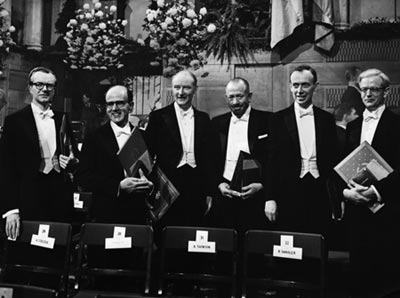
These two foundation stones of modern biology and medicine – DNA structure and protein structure – were recognized in the same year (1962) by the award of a Nobel Prize in Physiology or Medicine to Watson, Crick and Wilkins, and a Nobel Prize in Chemistry to Perutz and Kendrew. Franklin had died of cancer in 1958 at the tragically young age of just 37.
A particularly surprising feature of these discoveries was that they were both made in the Cavendish Laboratory – a physics laboratory. Under J J Thomson and then Ernest Rutherford, the Cavendish had played a dominant role in the development of atomic and nuclear physics in the years before the Second World War. But why had work in the then very new field of X-ray analysis of biomolecular structure reached such an advanced state in a physics laboratory?
The answer lies in two directions. The more obvious is the strength of experimental physics in Cambridge, beginning in the later part of the 19th century with Maxwell, Rayleigh and Thomson. This provided the intellectual environment where William and Lawrence Bragg (father and son) were trained, and where Lawrence Bragg – as an undergraduate and then as a research student – had the first ideas in 1912 that led them to invent the technique of X-ray structural analysis.
Although the diffraction of X-rays by crystals had already been discovered by Max von Laue, Walter Friedrich and Paul Knipping, it was Lawrence Bragg who first realized the simple way in which it could be understood. This was in terms of reflection by “Bragg planes” – sheets of atoms in different crystallographic directions that can diffract strongly at specific angles determined by the separation between sheets. Using this approach, the two Braggs were able to calculate the exact arrangement of sodium and chloride atoms in a crystal of salt. For this work, Lawrence Bragg, aged just 24, shared the Nobel Prize for Physics with his father in 1915. Both went on to play powerful roles in establishing major schools of X-ray analysis in Britain, building on their initial discovery and the many subsequent contributions that they made.
But while these earlier events created the right environment from which the 1953 discoveries could flow, that they actually did so depended on the personalities and decisions of many individuals, on many chance encounters, and on fortuitous sets of circumstances. As so often is the case in life, the outcome could have taken quite different directions on many occasions.
The early years: William Bragg, J D Bernal and Max Perutz
William Bragg graduated in mathematics in Cambridge in 1884, and immediately became professor of physics in Adelaide, Australia. In 1909 he returned to Britain to take up a chair in Leeds, where he continued his work on the nature of X-rays. He became director of the Royal Institution in London in 1923, where he attracted some outstanding young scientists interested in the X-ray field. Among them were William Astbury and John Desmond Bernal – both recent Cambridge graduates. They became interested in the problem of protein structure – Astbury as a result of being asked by Bragg to provide X-ray diagrams of wool and silk for lectures that he was to give.
Bernal moved back to Cambridge as a lecturer in structural crystallography in 1927, working in four dilapidated rooms that were later demolished to make way for the Austin wing of the Cavendish. In 1931 he was promoted to assistant director of research in crystallography, which was by then a sub-department of the Cavendish, although his group remained housed in the same old rooms as before. Bernal’s main scientific interest was initially in the atomic structure of crystals of metals and minerals, then of hormones and sterols, and of some amino acids – the building blocks of proteins.
Astbury, meanwhile, moved to Leeds in 1928, where he also began working on amino acids and proteins. He and Bernal corresponded amicably about Astbury’s unsuccessful attempts to obtain well-ordered X-ray diffraction patterns from crystals of the protein pepsin, and about the possibility of Bernal providing help in obtaining crystals of other proteins. In the event, a friend of Bernal’s in Cambridge called Glenn Millikan – son of Robert Millikan of oil-drop fame – happened to visit a lab in Uppsala, Sweden, where large crystals of pepsin had just been obtained. Millikan, who knew of Bernal’s interest in proteins, brought some of the crystals back to Cambridge, still in their mother-liquor.
Bernal and Dorothy Crowfoot (later Hodgkin) initially obtained patterns of the crystals in the dry state, as Astbury had, with similar disappointing results. But when Bernal observed the crystals in a light microscope, he noticed that on drying they became disordered, as the large amount of water in the crystal lattice evaporated. The X-ray experiment was then repeated, but now with the crystal surrounded by its mother-liquor and sealed in a glass capillary. This time they obtained patterns with large numbers of crystalline reflections from the fully hydrated crystals, revealing a hexagonal lattice with a 67 Å spacing and a third axis that was too long to be measured accurately.
This was the first defining moment in protein crystallography associated with the Cavendish. The results were published as a letter in Nature in 1934 (133 794), together with a related letter by Astbury (133 795). Astbury had deduced the presence of polypeptide chains – indicated by 4.5 Å and 10 Å rings that he had seen in diffraction patterns from dried crystals of pepsin. Subsequently, Astbury continued to pursue his pioneering studies of polypeptide-chain configurations in fibrous proteins. He also obtained the first X-ray patterns of partially oriented samples of DNA, showing the characteristic 3.4 Å axial repeat, which he correctly ascribed to the repeat of certain chemical structures (known as bases) along the polynucleotide chains.
In 1935 Max Perutz, a chemistry graduate from Vienna who wanted to do research on the structure of proteins, arrived in Cambridge to work as a graduate student with Bernal, to whom he had been recommended by his mentor in Austria. Despite the run-down appearance of the laboratory, Perutz found it a magical place to work, largely because of Bernal’s charismatic character. Bernal had a very wide range of interests, making contributions of great originality and force on everything from liquids, minerals, metals and organic compounds to proteins and viruses. He developed apparatus for X-ray data collection and contributed to the International Tables used in crystallographic data analysis. He wrote and lectured widely on science and society, and was well versed in history. During the Second World War he was one of the founders of “operational research”, and, as Louis Mountbatten’s scientific advisor, played a large part in the planning of the Normandy landings. Mountbatten described him as being “one of the most engaging personalities I have ever met…with a clear analytical brain…tireless and outspoken”. He was also a convinced and very active Marxist.
In 1936 Perutz was given excellent crystals of haemoglobin by Gilbert Adair, and soon produced the best X-ray diffraction patterns to date, with reflections extending out to a resolution of 2-3 Å. These were published jointly with Isadore Fankuchen in Nature in 1938, together with similarly promising X-ray patterns from crystals of the enzyme chymotrypsin. However, the observable diffraction pattern – i.e. the intensities and positions of the individual reflections – represents only half of the data needed to deduce the structure of the diffracting object. In mathematical terms, it gives the amplitude – but not the phases – of the terms in the 3D Fourier series that represents the object. Without the phase information, the data could not be deciphered and the atomic positions could not be determined.
With simpler structures made up of small numbers of atoms, where chemistry could provide considerable guidance as to the atomic arrangements, a solution could often be found by an educated trial-and-error process. But proteins, which contain thousands of atoms, were far too complicated for this to work. So despite the enormous amount of excellent data that could be (and was) collected, the solution remained tantalizingly out of reach.
One possibility that was considered at the time involved attaching a strongly scattering atom, such as gold or mercury, to a specific site on a protein in a crystal. The atom might produce changes in the intensities that could then be used to obtain the phase information. However, this solution was not pursued after preliminary experiments proved unpromising. The technical difficulties were great, and the belief grew that proteins had so many atoms that the intensities of the reflections would not be altered measurably by adding a single, heavy-metal atom. But the faith remained that detailed information about protein structure could be obtained from the X-ray patterns in some way, if only it could be discovered.


