Conventional ultrasound imaging – as routinely employed in hospital scans – works in reflection mode and provides qualitative images of soft tissue reflectivity, or echogenicity. In ultrasound tomography (UST), by contrast, many more measurements are taken and a computational algorithm is used to reconstruct quantitative images of acoustic properties, most commonly the sound speed.
Although UST was first proposed several decades ago, recent advances in hardware, not least in computational power, have led to a revival of interest and progress in the technique, in particular for breast imaging.
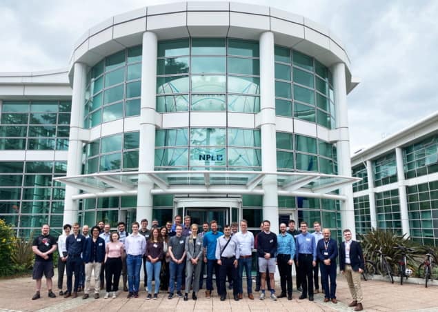
Bringing together UST research groups from around the globe, the 3rd International Workshop on Medical Ultrasound Tomography, MUST 2022, took place last month from 27–29 June, hosted by the National Physical Laboratory (NPL) in collaboration with Imperial College London and University College London.
The talks at the workshop covered all aspects of UST, from hardware design, through image reconstruction, to application in the clinic. But there were perhaps two dominant themes: breast imaging and the use of full-waveform inversions for image reconstruction.
The workshop was opened by NPL’s Chief Scientist, JT Janssen, who described some of the illustrious history of NPL and its current roles in maintaining standards and thereby accelerating research and innovation and facilitating trade.
The first invited speaker was Jeroen Veltman, a breast radiologist from the University of Twente in the Netherlands, who gave a clear description of the clinical workflow requirements and unmet needs in breast radiology. Recently, UST scanners developed by both QT Imaging and Delphinus Medical Technologies have been approved by the US Food and Drug Administration for breast imaging, and the conference attendees heard from the chief scientists behind both these scanners, James Wiskin and Neb Duric.
The group led by Nicole Ruiter, from Karlsruhe Institute of Technology, is a long-time pioneer of fully 3D breast UST technology. Ruiter spoke about UST technical challenges and system design, including presenting details of the group’s latest breast scanner.
The topic of image reconstruction using full-waveform methods was headlined by Jeroen Tromp from Princeton University, who gave a beautifully illustrated description of the state of the field of waveform tomography in the geosciences and seismology, and noted the considerable overlaps with biomedical imaging.

NPL launches Metrology for Medical Physics Centre
Continuing this theme, there were several talks concerned with applying UST to imaging the brain through the intact skull. The skull is a major barrier to ultrasound, so this represents a considerable challenge. It will be exciting to see, at the next MUST conference, how much progress has been made.
The meeting was supported by NPL, Precision Acoustics, Blatek, the Department for Business Energy & Industrial Strategy, and the UK Acoustics Network (UKAN), who sponsored an early career researcher prize for the best poster. The winner of the UKAN prize was Martin Angerer from Karlsruhe Institute of Technology, for his poster: A new generation of transducer arrays for 3D USCT III.


