The Acoustics 2023 Sydney conference, co-hosted by the Acoustical Society of America and the Australian Acoustical Society, brought together acousticians, researchers, musicians and other experts from around the world to share the latest developments in the field. Several of the presented studies described innovative applications of acoustics in healthcare, including the use of acoustic cavitation for needle-free vaccine delivery, and a wearable ultrasound transducer that tracks muscle dynamics during recovery from injury.
Ultrasound enables painless vaccination
Darcy Dunn-Lawless from the University of Oxford’s Institute of Biomedical Engineering described the use of ultrasound for needle-free delivery of vaccines.
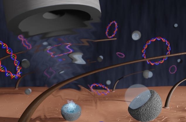
Aiming to circumvent the fear of needles suffered by many adults, and many more children, Dunn-Lawless and colleagues are exploiting an acoustic effect called cavitation, in which a sound wave causes the formation and popping of bubbles. When these bubbles collapse, they release a concentrated burst of mechanical energy.
The idea is to use these energy bursts in three ways: to clear passages through the outer layer of dead skin cells and allow vaccine molecules to pass through; to actively force vaccine molecules into the body; and to open up cell membranes inside the body. To enhance the cavitation activity, the researchers employed nanometre-sized particles called protein cavitation nuclei (PCaNs) – essentially cup-shaped protein particles – to support the gas bubbles.
In tests on mice, the researchers compared the immune response generated by standard intradermal vaccination of a DNA vaccine versus the cavitation approach. For cavitation-based delivery, they mixed PCaNs with the DNA vaccine in a chamber placed on the animal’s skin and exposed to ultrasound for two minutes.
They found that conventional injection delivered several orders of magnitude more vaccine molecules than the cavitation approach. “However, this is where things get interesting,” explained Dunn-Lawless at a press conference. “When you look at the immune response generated by both of these delivery methods, the antibody concentration, you can see that the cavitation group received a significantly higher immune response, even though they received so many fewer molecules of vaccine.”
He noted that this a particularly exciting result, firstly as it confirms that it’s possible to deliver vaccines in this way. But also because it shows that the needle-free technique can, in theory, allow the body to achieve greater immune response with less vaccine, making vaccination more efficient.
The mechanism underlying this effect is not yet clear, but Dunn-Lawless suggested that it may be due to cavitation activity opening up cell membranes and allowing molecules into the cells. Or in other words, although fewer molecules get into the body, the ones that do, get into the correct place. This could be particularly favourable for DNA vaccines, which are currently difficult to deliver as they need to get inside the cell to function.
Monitoring muscle recovery in real time
Recovery from musculoskeletal injury can be a long and difficult process. It’s therefore important to track a patient’s progress as they undergo rehabilitation and slowly rebuild muscle strength. But direct measures of muscle function during physical activity are not readily available, and few medical technologies can be used while the patient is moving, which can hinder treatment and rehabilitation.
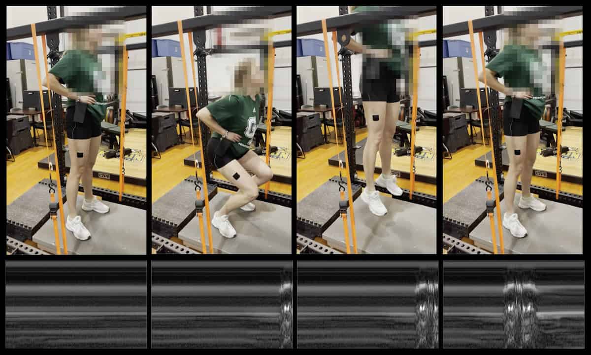
One option is ultrasonography, which can provide non-invasive images of tissue under the skin and reveal how different muscle groups move and contract during dynamic physical activity. Traditional ultrasound systems, however, are large and cumbersome, require the patient to be tethered to the instrument, and are thus not conducive to real-time imaging during activity.
So Parag Chitnis from George Mason University and colleagues decided to build their own ultrasound device from scratch. They designed a compact wearable ultrasound system that moves with the patient and produces clinically relevant information about muscle function during physical activity.
To do this, the researchers developed new ultrasound technology that relies on the transmission of low-voltage, long-duration chirps – as opposed to the conventionally used very high-voltage, short-duration pulse sequences. This enabled them to employ low-cost electronic components, like those found in a car radio, to design a simpler, portable ultrasound system that could be powered by batteries and attached to a patient. They call the new approach SMART-US, or simultaneous musculoskeletal assessment with real-time ultrasound.
The team tested the approach on a subject performing counter movement jumps (a routine exercise for evaluating the health and function of lower limbs and knee joints) on a force plate with an ultrasound transducer attached to their leg. The SMART-US device provided real-time feedback on the level of muscle activation and function during the jumps, with significant correlation seen between force data and ultrasound measurements. Chitnis added that the technique can also be used to examine several different muscles simultaneously.

Wearable ultrasound sensor provides continuous cardiac imaging
“Ultrasound-based biofeedback can help personalize therapy and rehabilitation to improve treatment outcomes,” he explained at a press conference. “Other applications that we envision for our technology include personal fitness, athletic training and sports medicine, military health, stroke rehabilitation and assessing the risk of falls in elderly populations.”
The next goal is technology transfer, to put the device through FDA clearance so the team can perform clinical studies for rehabilitation. Moving forward, Chitnis envisages that clinics would be able to purchase a basic-level system for just a few hundred dollars.
