Imaging plays a vital role in diagnosing brain disease and disorders, as well as advancing our understanding of how the human brain works. Existing brain imaging modalities, however, usually require the subject to lie flat and motionless – precluding use in people who cannot remain still or studies of the brain in motion.
To address these limitations, neuroscientists at West Virginia University have developed a wearable, motion-compatible brain positron emission tomography (PET) imager and demonstrated its use in a real-world setting. The device, described in Communications Medicine, could potentially enable previously impossible neuroimaging studies.
“We wanted to create and test a tool that could grant access to imaging the brain – including deep areas – while humans are moving around,” explains senior author Julie Brefczynski-Lewis. “We hope our device could allow the investigation of research questions related to natural upright behaviours, or the study of patients who are normally sedated due to movement issues or challenges in understanding the need to be perfectly still for a scan, which could happen with cognitive impairments or dementias.”
PET scans provide information on neuronal and functional activity by imaging the uptake of radioactive tracers in the brain. But clinical PET systems are extremely sensitive to motion and require supine (lying down) imaging and dedicated scanning rooms. There are neuroimaging techniques that can be used with patients upright and moving – such as functional near-infrared spectroscopy and high-density diffuse optical tomography – but these optical approaches only image the brain surface. Activity in deep brain structures remains unseen.

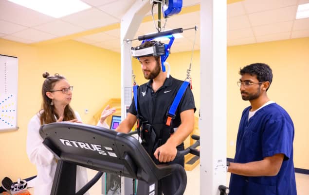
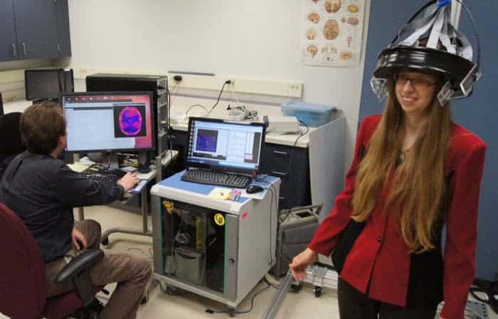
To enable upright and motion-tolerant imaging, Brefczynski-Lewis and colleagues designed the AMPET (ambulatory motion-enabling positron emission tomography), a helmet-like device that moves along with the subject’s head. The imager is made from a ring of 12 lightweight detector modules, each comprising arrays of silicon photomultipliers coupled to pixelated scintillation crystal arrays. The imager ring has a 21 cm field-of-view and a central spatial resolution of 2 mm in the tangential direction and 2.8 mm in the radial direction.
Real-world scenarios
The researchers tested the AMPET device on 11 volunteer patients who were scheduled for a clinical PET scan on the same day. The helmet was positioned the on the participant’s head such that it imaged the top of the brain, comprising the primary motor areas. Although it only weighs 3 kg, the team chose to suspend the helmet from above so that participants would not feel any weight while moving their head.
Patients received a low dose (10–20% of their total prescription) of the metabolic PET tracer 18F-FDG. “We chose a very low dose that was within the daily dose for clinical patients,” says Brefczynski-Lewis. “Some applications may require a slightly higher dose for extra sensitivity, but because the detectors are so close to the head, a full clinical-like dose would not likely be necessary.”
Immediately after tracer injection, each participant underwent AMPET imaging for 6 min while they switched between standing still and walking-in-place every 30 s. Following a 5 min transition, subjects were then scanned for another 5 min while alternating between sitting still and lifting their leg while seated. In this second imaging session, the team moved the AMPET lower around the head for five participants, to image deeper brain structures.
Meeting the goals
The team defined three goals to validate the AMPET prototype: motion artefacts of less than 2 mm; differential activation of cortical regions of interest (ROIs) related to leg movement; and differential activation to walking movements in deep brain structures.
The walking versus standing task allowed the researchers to test for any motion of the imager relative to the head. They observed an average movement-related misalignment of just 1.3 mm. Analysis of task-related activity showed the expected brain image patterns during walking, with activity in ROIs that control leg movements significantly greater than in all other imaged ROIs.
In four participants where activity was measured from deep brain structures (the fifth had incorrect helmet placement), the team observed differential activation in various deep lying structures, including the basal nuclei.
The researchers note that one volunteer had a prosthetic right leg. While performing upright walking, his brain patterns showed greater metabolic activity in the area that represented the intact leg. In contrast, no difference in activity between left and right leg ROIs was measured in the other participants.
Portable brain scanner allows PET in motion
Brefczynski-Lewis tells Physics World that patients found the AMPET reasonably comfortable and did not feel its weight on their head or neck. Certain movements, however, were slightly inhibited, especially tilting the head towards the shoulders. “Our engineer collaborators recommended a gyroscope mechanism to enable free movement in all directions,” she says.
As well as validating the prototype, the study also identified upgrades required for the AMPET and similar systems. “The great thing about a real-world study on humans was that it showed us which logistics to optimize,” explains Brefczynski-Lewis. “We are developing a system for good placement and monitoring the alignment of the imager relative to the head, as well as widening the coverage to increase sensitivity, and testing a movement task using a bolus-infusion paradigm.”


