Panicos Kyriacou is chief scientist at the UK-based start-up Crainio, which has developed an inexpensive light-based instrument that enables non-invasive measurement of intracranial pressure. He tells Tami Freeman that the sensor could improve the diagnoses of traumatic brain injury
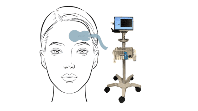
Traumatic brain injury (TBI), caused by a sudden impact to the head, is a leading cause of death and disability. After such an injury, the most important indicator of how severe the injury is intracranial pressure – the pressure inside the skull. But currently, the only way to assess this is by inserting a pressure sensor into the patient’s brain. UK-based startup Crainio aims to change this by developing a non-invasive method to measure intracranial pressure using a simple optical probe attached to the patient’s forehead.
Can you explain why diagnosing TBI is such an important clinical challenge?
Every three minutes in the UK, someone is admitted to hospital with a head injury, it’s a very common problem. But when someone has a blow to the head, nobody knows how bad it is until they actually reach the hospital. TBI is something that, at the moment, cannot be assessed at the point of injury.
From the time of impact to the time that the patient receives an assessment by a neurosurgical expert is known as the golden hour. And nobody knows what’s happening to the brain during this time – you don’t know how best to manage the patient, whether they have a severe TBI with intracranial pressure rising in the head, or just a concussion or a medium TBI.
Once at the hospital, the neurosurgeons have to assess the patient’s intracranial pressure, to determine whether it is above the threshold that classifies the injury as severe. And to do that, they have to drill a hole in the head – literally – and place an electrical probe into the brain. This really is one of the most invasive non-therapeutic procedures, and you obviously can’t do this to every patient that comes with a blow in the head. It has its risks, there is a risk of haemorrhage or of infection.
Therefore, there’s a need to develop technologies that can measure intracranial pressure more effectively, earlier and in a non-invasive manner. For many years, this was almost like a dream: “How can you access the brain and see if the pressure is rising in the brain, just by placing an optical sensor on the forehead?”
Crainio has now created such a non-invasive sensor; what led to this breakthrough?
The research goes back to 2016, at the Research Centre for Biomedical Engineering at City, University of London (now City St George’s, University of London), when the National Institute for Health Research (NIHR) gave us our first grant to investigate the feasibility of a non-invasive intracranial sensor based on light technologies. We developed a prototype, secured the intellectual property and conducted a feasibility study on TBI patients at the Royal London Hospital, the biggest trauma hospital in the UK.
It was back in 2021, before Crainio was established, that we first discovered that after we shone certain frequencies of light, like near-infrared, into the brain through the forehead, the optical signals coming back – known as the photoplethysmogram, or PPG – contained information about the physiology or the haemodynamics of the brain.
When the pressure in the brain rises, the brain swells up, but it cannot go anywhere because the skull is like concrete. Therefore, the arteries and vessels in the brain are compressed by that pressure. PPG measures changes in blood volume as it pulses through the arteries during the cardiac cycle. If you have a viscoelastic artery that is opening and closing, the volume of blood changes and this is captured by the PPG. Now, if you have an artery that is compromised, pushed down because of pressure in the brain, that viscoelastic property is impacted and that will impact the PPG.
Changes in the PPG signal due to changes arising from compression of the vessels in the brain, can give us information about the intracranial pressure. And we developed algorithms to interrogate this optical signal and machine learning models to estimate intracranial pressure.
How did the establishment of Crainio help to progress the sensor technology?
Following our research within the university, Crainio was set up in 2022. It brought together a team of experts in medical devices and optical sensors to lead the further development and commercialization of this device. And this small team worked tirelessly over the last few years to generate funding to progress the development of the optical sensor technology and bring it to a level that is ready for further clinical trials.
In 2023, Crainio was successful with an Innovate UK biomedical catalyst grant, which will enable the company to engage in a clinical feasibility study, optimize the probe technology and further develop the algorithms. The company was later awarded another NIHR grant to move into a validation study.
The interest in this project has been overwhelming. We’ve had a very positive feedback from the neurocritical care community. But we also see a lot of interest from communities where injury to the brain is significant, such as rugby associations, for example.
Could the device be used in the field, at the site of an accident?
While Crainio’s primary focus is to deliver a technology for use in critical care, the system could also be used in ambulances, in helicopters, in transfer patients and beyond. The device is non-invasive, the sensor is just like a sticking plaster on the forehead and the backend is a small box containing all the electronics. In the past few years, working in a research environment, the technology was connected into a laptop computer. But we are now transferring everything into a graphical interface, with a monitor to be able to see the signals and the intracranial pressure values in a portable device.
Following preliminary tests on patients, Crainio is now starting a new clinical trial. What do you hope to achieve with the next measurements?
The first study, a feasibility study on the sensor technology, was done during the time when the project was within the university. The second round is led by Crainio using a more optimized probe. Learning from the technical challenges we had in the first study, we tried to mitigate them with a new probe design. We’ve also learned more about the challenges associated with the acquisition of signals, the type of patients, how long we should monitor.
We are now at the stage where Crainio has redeveloped the sensor and it looks amazing. The technology has received approval by MHRA, the UK regulator, for clinical studies and ethical approvals have been secured. This will be an opportunity to work with the new probe, which has more advanced electronics that enable more detailed acquisition of signals from TBI patients.
We are again partnering with the Royal London Hospital, as well as collaborators from the traumatic brain injury team at Cambridge and we’re expecting to enter clinical trials soon. These are patients admitted into neurocritical trauma units and they all have an invasive intracranial pressure bolt. This will allow us to compare the physiological signal coming from our intracranial pressure sensor with the gold standard.
The signals will be analysed by Crainio’s data science team, with machine learning algorithms used to look at changes in the PPG signal, extract morphological features and build models to develop the technology further. So we’re enriching the study with a more advanced technology, and this should lead to more accurate machine learning models for correctly capturing dynamic changes in intracranial pressure.
The primary motivation of Crainio is to create solutions for healthcare, developing a technology that can help clinicians to diagnose traumatic brain injury effectively, faster, accurately and earlier
This time around, we will also record more information from the patients. We will look at CT scans to see whether scalp density and thickness have an impact. We will also collect data from commercial medical monitors within neurocritical care to see the relation between intracranial pressure and other physiological data acquired in the patients. We aim to expand our knowledge of what happens when a patient’s intracranial pressure rises – what happens to their blood pressures? What happens to other physiological measurements?
How far away is the system from being used as a standard clinical tool?
Crainio is very ambitious. We’re hoping that within the next couple of years we will progress adequately in order to achieve CE marking and all meet the standards that are necessary to launch a medical device.
The primary motivation of Crainio is to create solutions for healthcare, developing a technology that can help clinicians to diagnose TBI effectively, faster, accurately and earlier. This can only yield better outcomes and improve patients’ quality-of-life.
Of course, as a company we’re interested in being successful commercially. But the ambition here is, first of all, to keep the cost affordable. We live in a world where medical technologies need to be affordable, not only for Western nations, but for nations that cannot afford state-of-the-art technologies. So this is another of Crainio’s primary aims, to create a technology that could be used widely, because there is a massive need, but also because it’s affordable.
- To hear the full interview with Panicos Kyriacou listen to our Physics World Weekly podcast
