Physicists in France have generated supersonic sound waves in human tissue for the first time by exploiting the acoustic equivalent of Cerenkov radiation – the light emitted by charged particles when they travel through a medium faster than the speed of light in that medium. Mathias Fink and colleagues at the Laboratoire Ondes et Acoustiques in Paris say that their results could have applications for ultrasound imaging in medicine (J Bercoff et al. 2004 Appl. Phys. Lett 84 2202).

Ultrasound is best known for providing the first images of unborn babies in the womb but it also widely used in many other medical applications. Most ultrasound machines convert an electrical signal directly into mechanical vibrations through the piezoelectric effect. However, these devices produce longitudinal sound waves and only operate within a narrow range of megahertz frequencies. Shear sound waves – which could provide more information about the object being scanned – cannot be used at these frequencies because they are absorbed by human tissue.
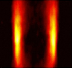
Now, Fink and colleagues have overcome this problem by generating shear sound waves at sonic frequencies. They focus an ultrasound beam in the tissue being studied to create a local vibration that acts as a source of further shear waves. This source can be moved by focusing the original ultrasound wave at different depths in the tissue, and if the source is made to move faster than the speed of shear waves in the tissue, the beam can reach supersonic speeds in just tens of milliseconds. The waves – which initially propagate in a so-called Mach cone – are distorted by any inhomogeneities they encounter in the tissue, and these distortions are analysed to produce an image (see figures).
“Our ultrafast echographic device is the only prototype in the world that is able to generate a supersonic regime and also image the resulting shear waves that propagate in the body,” Fink told PhysicsWeb. “This system is able to compute 5000 ultrasound images per second, which is more than 100 times faster than conventional ultrasound techniques.”
Fink says the technique has already been successfully tested on healthy human breast tissue and could prove useful in diagnosing cancer. His team now plans to test the method on patients with tumours.