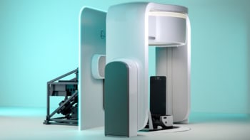
Multimodal system delivers photon and particle beams
ViewRay has developed a multimodal treatment system that can deliver radiation therapy via a photon beam and also via a particle beam without having to move the patient (WO/2019/112880). The system can also include an MRI device to acquire patient images during treatment. These patient MRI data can be used to perform real-time calculations of the location of dose deposition for the particle beam and for the photon beam, taking into account the influence of the magnetic field on the beams, as well as interactions with soft tissues. The MRI data and beam information can be used to calculate accumulated dose deposition during radiotherapy. Optionally, the treatment can be re-optimized based on this calculated dose deposition.
Intelligent system offers automated radiotherapy planning
An intelligent automatic radiotherapy planning system is described by Samsung Life Public Welfare Foundation of Korea (WO/2019/143120). The method involves creating a database containing personal, diagnostic and disease stage information, plus medical images, for existing patients who have undergone radiotherapy. It also includes radiotherapy dose and irradiation data for the patients, and their clinical results following radiotherapy. The system generates a plan prediction model by performing artificial intelligence-based correlation and regression analysis on these data. It then uses this prediction model, along with personal, diagnostic and imaging data, to create a radiotherapy plan for a new patient. After delivering this radiotherapy plan to the new patient, their plan and outcome data are then added to the database.
IORT device reduces scatter radiation
Intra-operative radio therapy (IORT), irradiating the tumour bed during cancer surgery, has become established in recent years due to the development of mobile accelerators. One crucial aspect of an IORT accelerator is the quantity of scatter radiation produced during treatment. SIT of Italy has invented an IORT device that minimizes scatter radiation in the entire surrounding space, whilst maintaining dimensions compatible with a standard operating room (WO/2019/142217). The system comprises: a source of particles; an accelerating device that directs the particle beam onto a target through an applicator; a scattering filter that keeps the distance between the particle source and the target suitable for use in an operating room; and an optical system for collimating the particle beam. This collimating system, which is placed between the filter and the applicator, includes a primary screen to shield radiation produced by the scattering filter, a secondary screen to shield the photons produced on the primary screen and a collimating apparatus for housing the monitor chambers.
Apparatus acquires PET data during hadron therapy
Researchers at Università di Pisa and INFN have devised techniques for acquiring PET data for in-beam monitoring during hadron therapy (WO/2019/138384). The apparatus comprises detectors that detect radiation emitted by the irradiated target and a data processing system that reconstructs PET data and determines the beam range. The data processing system determines a time distribution of the coincidence event rate acquired by a detector and then samples each spill interval at a gigahertz frequency to obtain a fine-grained event rate distribution. For each spill interval, the processor analyses frequencies of the fine-grained distribution to find an initial frequency that’s compatible with the RF signal of the hadron beam, uses this initial frequency to estimate the period of the micro-bunches and transforms time values of coincidence events into phase values, based on the estimated period of the micro-bunches. For each micro-bunch, the processor filters out coincidence events associated with background noise from the hadron beam and keeps the remaining events.
Ultrasound enhances drug uptake into cancer cells
A system for inducing sonoporation of drug-carrying liposomes into a tumour, to enhance drug uptake into the targeted cancer cells, is detailed by Freedom Waves (WO/2019/123411). The system includes a generator that provides electrical energy at an ultrasound frequency. An ultrasound probe connected to the generator converts this electrical energy into low-intensity pulsed ultrasonic waves, defined by operation parameters such as frequency, duty cycle and ultrasound operation time. An input device enables the operator to enter configuration data, including the tumour type and grade, drug type, location of secondary tumour and body measurements. A processor then determines the operation parameters on the basis of these input data and controls the generator and ultrasound probe to operate according to the determined values.


