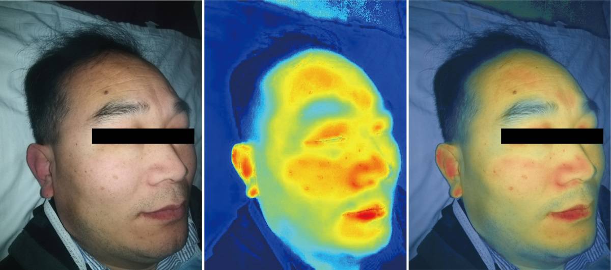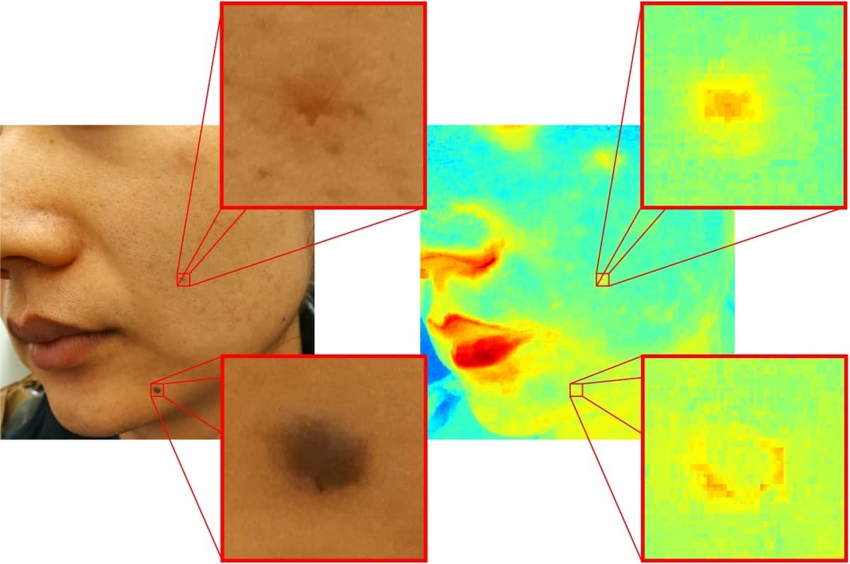
The built-in RGB camera in a modern smartphone can be used to create a hyperspectral imaging system for analysis and monitoring of skin features. Ruikang Wang and Quinghua He from the University of Washington suggest that the ability to produce images comparable to those from expensive hyperspectral imaging systems may eventually enable widespread use of smartphone-based hyperspectral imaging in low-resource settings and rural areas (Biomed. Opt. Express 10.1364/BOE.378470).
In a hyperspectral image, each pixel contains information regarding a series of narrow wavelength bands. Hyperspectral imaging can be used to determine levels of chromophores with skin tissue – such as haemoglobin and melanin, for example – generating data that help differentiate melanomas from pigmented skin lesions. Variation in melanin may be seen in some skin cancers, nevus and skin pigmentation, while haemoglobin concentration may indicate vascular abnormalities and inflammation.
Hyperspectral imaging systems have been in clinical use for decades, but have a complex design, are expensive and generally limited to use in clinical laboratories. Today’s smartphones typically incorporate RGB cameras with 8 to 12 million pixels and are capable of high-speed photography. To exploit this capability, Wang and He applied Wiener estimation to transform RGB images captured by smartphone cameras into “pseudo”-hyperspectral images with 16 wavebands covering 470–620 nm. They processed the reconstructed hyperspectral images using weighted subtractions between wavebands to extract absorption information caused by specific chromophores, such as haemoglobin or melanin, within skin tissue.
The researchers captured images from two volunteers with redness and moles on their facial skin. They also acquired images in the dark, using the smartphone camera’s built-in flashlight or a fluorescent lamp as illumination sources. Both light sources worked equally well, demonstrating flexibility in terms of using different illumination conditions.

As blood vessels are localized within relatively deep skin tissue, light with a longer penetration depth is suitable for detection. To extract spatial haemoglobin absorption information, the researchers therefore applied weighted subtractions between green and red wavebands. Melanin, on the other hand, exists in superficial skin layers, so they extracted melanin absorption data using weighted subtractions between blue and green bands.
Comparing the melanin absorption data with results from a snapshot hyperspectral camera showed that the absorption map created from the smartphone exhibited much better image resolution, because the smartphone camera has many more pixels than the snapshot camera.
Wang and He also examined whether it is possible to evaluate heart rate from a time series of blood information maps, by monitoring changes in blood absorption intensity in the skin. Using a fixed support to keep facial skin stable, they recorded a smartphone video under flashlight illumination.
They extracted the blood absorption map from every frame in the video and summed the signals for each frame. By Fourier transforming the temporal data, they created a plot in the frequency domain and identified a main frequency peak around 1.05 Hz. This matched the 1.05 Hz heart-beat frequency recorded by a pulse sensor for reference.
The researchers also tested the smartphone’s ability to monitor vascular occlusion, by recording images of a volunteer’s finger with pressure from a rubber ring applied for 60 s to create a vascular occlusion. The smartphone video recorded this skin vascular occlusion, as well as restoration of the finger to normal state.

Three-in-one approach detects early skin cancers
“Compared with conventional hyperspectral imaging systems, which mostly rely on lasers or tunable optical filters, the smartphone-based hyperspectral imaging system eliminates the internal time difference within frames, greatly improving the imaging speed and immunity to motion artefacts,” the researchers write. “Most importantly, our strategy does not require any modification or addition to the existing smartphones, which makes hyperspectral imaging and analysis of skin tissue possible in daily scenes out of labs.”
“In addition to future clinical applications, we envision that smartphone imaging could provide excellent impact on cosmetic consumers and also for the cosmetic industry,” comments Wang.
Wang and He tell Physics World that they are currently developing a smartphone-based app to provide information about blood perfusion, pigmentation, and porphyrin-containing bacteria and collagen content of the skin. They are also currently enrolling a mix of volunteers with various skin colours to check whether there is any skin-colour dependence in the results from their hyperspectral imaging system.


