Vascular leakage is an important biomarker for assessing vision-threatening retinal diseases such as age-related macular degeneration and diabetic retinopathy. Fluorescence angiography, the imaging exam currently used to identify vascular leakage, lacks depth resolution, which can hamper the identification and precise localization of leaky blood vessels. Optical coherence tomography (OCT) is a newer clinical ophthalmic imaging technology that provides high-resolution images and rapid volumetric scanning. To date, however, OCT has been unable to visualize vascular leakage.
Researchers at the Medical University of Vienna are developing a new OCT method, exogenous contrast-enhanced leakage OCT (ExCEL-OCT), which measures the diffusion of tracer particles around leaky vasculature. Writing in Biomedical Optics Express, they describe the use of an OCT contrast agent to visualize the slow extravasation of tracer particles from leaky blood vessels in laboratory mice. In just a single scan, ExCEL-OCT provided high-resolution structural, angiographic and leakage information that was spatially and temporally co-registered, and separable.
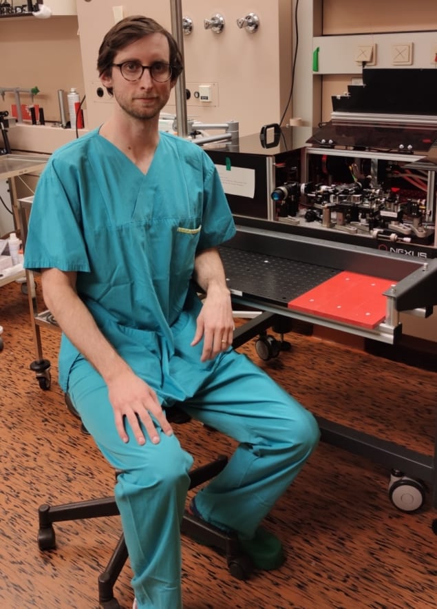
The researchers used a custom-built OCT ophthalmoscope designed for rodent eye imaging. For the contrast agent, they selected Intralipid 20%, an emulsion of lipid particles that can dramatically improve OCT intensity and angiogram signals.

Principal investigators Bernhard Baumann and Conrad Merkle, of the Center for Medical Physics and Biomedical Engineering, and colleagues imaged the eyes of mice with leaky retinal vasculature and control mice. To track the leakage of tracer particles, they performed multiple angiogram scans (using a traditional 3D angiography protocol covering a 1 x 1 mm field-of-view centred on the optic disc) before and after injection of the OCT contrast agent, using the data to generate both angiogram and leakage maps.
The researchers employed post-processing to compensate for motion and flatten the retina. They also developed novel data processing methods to highlight the scattering signal from the extravasated Intralipid particles. To discriminate between the various signals, they used selective decorrelation gates.
“The key idea is that OCT signals in static tissue will decorrelate slowly because the tissue is not moving,” the researchers explain. “Signal within vessels will decorrelate rapidly due to the relatively high speed of the red blood cells and Intralipid particles passing through the voxel. Extravasated Intralipid particles, which are driven by slower diffusion processes rather than blood flow, will have a decorrelation rate that falls somewhere between the two.” As such, they expect much stronger extravasation and diffusion signal will be observed around leaky vessels.
The researchers used long interscan times to highlight diffusing tracer particles. They created the ExCEL signal by subtracting angiogram signals of different lag times, to specifically highlight leakage of different diffusion rates and remove the intravascular signal. They also created depth profiles, leakage maps and fly-through videos showing leakage over time.
The contrast agent dramatically increased the visibility of neovascularizations (new blood vessels) growing into a retinal lesion. Vascular leakage could be tracked over time, with results demonstrating a clear increase in ExCEL signal, visible as a circular or spherical bloom of signal, following administration of the contrast. The researchers confirmed this finding by blind grading 83 leakage and control volumes.
In addition to showing leakage in 3D, the researchers also demonstrated that for the most part, the leakage signal was separable from the angiogram signal, which is not the case for traditional fluorescence methods. Colour coding and overlaying the angiogram and leakage data revealed which blood vessels the leakage surrounds.
The researchers note that the current system produces a high number of false-positive leakage signals, and that they could not exclusively distinguish diffusion from slow intravascular flow or bulk motion of static tissue. As OCT systems become faster and motion compensation software improves, higher temporal resolution for ExCEL measurements and shorter interscan times may change this.
The team describe this work as “a starting point for future in vivo 3D volumetric leakage studies”, noting that the new method can be easily implemented in conventional OCT systems across the world.

Quantum-inspired detection method generates high-quality OCT images
“We hope to continue to improve our methods to the point where they can be used clinically,” Merkle tells Physics World. “To do this, we need to continue to develop the methods and post-processing to improve signal quality and reduce false-positive signals, investigate alternate sources of contrast, and/or improve the contrast agent to increase sensitivity and reduce dose. We are most interested in the first two options, as nanoparticle fabrication is something that other research groups are already investigating with great results.”
“Beyond this work on vascular leakage, we are also investigating hardware development of polarization-sensitive and visible light OCT systems, longitudinal studies of small-animal disease models, and ex vivo imaging of human brain tissue,” Merkle adds. “My own projects are currently focused on improving clinical OCT through software rather than hardware. Our hope is that by working within the limitations of existing clinical systems, we can develop methods to improve diagnostics at a larger scale and lower cost compared to hardware-based solutions that require new OCT systems.”


