
The epithelial–mesenchymal transition (EMT) is a process by which polarized epithelial cells undergo multiple biochemical changes enabling their transfer into a mesenchymal cell phenotype, which is characterized by an enhanced migratory capacity, invasiveness and elevated resistance to cell death. Although it is known that EMT plays an important role in tumour metastasis, the regulation mechanism is still to be elucidated.
Three-dimensional bioprinting has emerged as a useful tool for the study of cancer mechanisms, because it can mimic the in vivo microenvironment of tumour cells by the precise control of cells and biomaterials. Now, a team of researchers from Tsinghua University, Chinese Academy of Medical Sciences and Drexel University has implemented a 3D printed cervical tumour model to study the EMT process (Biofabrication 10 044102). Their findings provide a better understanding of the regulation mechanism of EMT, for use in future therapeutic programmes.
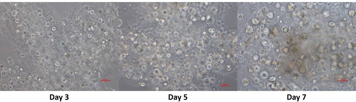
The researchers used HeLa cells (the oldest human cell line, derived from cervical cancer cells) to fabricate a cervical tumour model with a cuboid structure, with interconnected channels to facilitate sufficient transfer of nutrients, oxygen and metabolic waste. After printing, the cells proliferated and grew, confirming that the construct represents an advanced cancer model.
EMT can be induced or regulated by different factors and cytokines (small proteins that play an important role in cell signalling), with TGF-β (transforming growth factor beta) considered the major inducer of EMT in a variety of cell types and tissues.
As such, the authors evaluated the effect of TGF-β treatment on HeLa cells and their EMT phenotype by looking at the protein expression of target biomarkers like E-cadherin, together with snail (which shows the acquisition of a mesenchymal phenotype), vimentin and N-cadherin. The results indicated loss of epithelial markers and the acquisition of mesenchymal markers, suggesting that EMT was achieved in the 3D-printed HeLa cell construct.
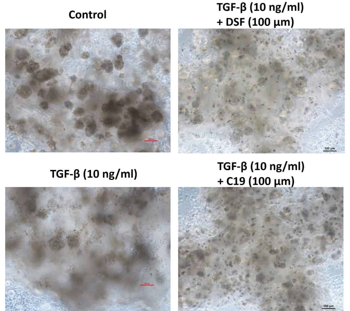
Further, to confirm that EMT was induced by TGF-β in the 3D printed cervical cancer model, the authors treated the 3D-printed HeLa cell construct with disulfiram (an anti-alcoholism drug) and the EMT pathway inhibitor C19, 24 hours before TGF-β induction. As expected, HeLa cells kept a more epithelial-like appearance after this treatment. The results of real-time PCR (polymerase chain reaction) indicated that the treatment with disulfiram and C19 inhibited the TGF-β-induced EMT process by modifying the expression of EMT-related proteins.
The authors have established a cervical tumour model by 3D printing of HeLa cells and achieved the EMT process by TGF-β treatment. These results may help researchers to better understand the regulation of cervical cancer metastasis.


