Researchers at Northwestern University have printed a 3D mini-tissue that mimics the bile duct. They achieved this by combining peptides amphiphiles, which form a scaffold, with bioink consisting of cells and growth factors (Biofabrication 10 035010).
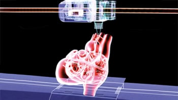
Three-dimensional (3D) bioprinting uses conventional 3D printing methods to mimic natural tissue states and promote self-assembly. 3D bioprinting uses cells and bioinks – natural or synthetic printable materials used with signal molecules such as growth factors and cytokines – to engineer tissue-like structures, or “mini-tissues”.
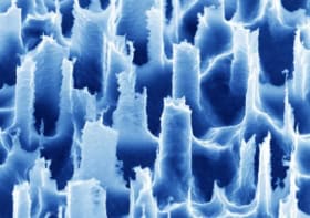
Cells in vivo need a fibrous scaffold that mimics the extracellular matrix (ECM), along with signalling molecules, to perform a desired function in a tissue. In bioprinting, the ECM is provided by a versatile class of peptide-based molecules known as peptides amphiphiles (PAs). PAs self-assemble into nanofibres and can be modified for different tissue engineering applications such as bone regeneration. Currently, the challenge is to develop and fine-tune bioinks that can be easily printed and meet the requirements for biomedical applications.
A stable bio-nanostructure
With this in mind, Ming Yan and colleagues have successfully 3D printed a nanostructure consisting of bioink, PAs and bile duct cells (cholangiocytes). They mixed thiolated-gelatin, PAs and cholangiocytes at 37°C and 3D printed the nanostructure at 4°C. The bioinks printed into filaments that retained integrity and could support multi-layered scaffolds. The bioink-PA scaffold was made stable by cross-linking a derivative of ethylene glycol with calcium ions. The scaffold remained stable for a long time (more than one month) in culture at 37 °C.
The researchers also investigated the influence on cholangiocytes of including a laminin-derived peptide (Ile-Lys-Val-Ala-Val, IKVAV) within the bioink. Laminin is an ECM molecule (found in the basement membrane) necessary for cell adhesion. After bioprinting, the cholangiocytes remained viable in vitro. Staining showed the formation of functional bile-cell-based tube structures, with enhanced morphology of these nanostructures observed when cultured in IKVAV-bioink. This is the first time that a bioink-based system supplemented with PAs has been used for a specific biological application – bile duct tissue engineering.
3D bioprinted scaffolds show promise in tissue engineering and regenerative medicine. For instance, complex spatial models need to be tested in order to bioprint a functional liver tissue with blood vessels, bile ducts and liver cells.
Building on the present study, the scientists now want to optimize the peptide concentration and test other signalling molecules within the bioinks to enhance the formation of functional tubular structures reminiscent of the architecture seen in natural liver. In addition to bioprinting, the PA-bioinks can help establish adaptable in vitro systems for modelling diseases such as bile duct cancer and discovering new drugs.


