Ultrasound is one of the most common medical imaging tools, but the electronic components in ultrasound probes make it difficult to miniaturize them for endoscopic applications. Such electronic ultrasound systems are also unsuitable for use within MRI scanners.
To address these shortcomings, researchers from University College London have developed an ultrasound system that uses optical, instead of electronic, components. The team has now demonstrated the first use of an all-optical ultrasound imager for video-rate, real-time 2D imaging of biological tissue (Biomed. Opt. Express 9 3481).
“All-optical ultrasound imaging probes have the potential to revolutionize image-guided interventions,” says first author Erwin Alles. “A lack of electronics and the resulting MRI compatibility will allow for true multimodality image guidance, with probes that are potentially just a fraction of the cost of conventional electronic counterparts.”
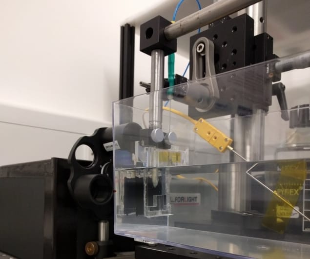
All-optical ultrasound systems eliminate the electronic transducers in standard ultrasonic probes by using light to both transmit and receive the ultrasound. Pulsed laser light generates ultrasound waves, scanning mirrors control the transmission of the waves into tissue, and a fibre-optic sensor receives the reflected waves.
The team also developed methods to acquire and display images at video rates. “Through the combination of a new imaging paradigm, new optical ultrasound generating materials, optimized ultrasound source geometries and a highly sensitive fibre-optic ultrasound detector, we achieved image frame rates that were up to three orders of magnitude faster than the current state-of-the-art,” Alles explains.
Optical components are easily miniaturized, offering the potential to create a minimally invasive probe. The scanning mirrors built into the device enable it to acquire images in different modes, and rapidly switch between modes without needing to swap the imaging probe. In addition, the light source can be dynamically adjusted to generate either low-frequency ultrasound, which penetrates deep into tissue, or high-frequency ultrasound, which offers higher-resolution images at a shallower depth.
The team tested their prototype system by imaging a deceased zebrafish, as well as an ex vivo pig artery manipulated to emulate the dynamics of pulsing blood. The all-optical device exhibited comparable imaging capabilities to an electronic high-frequency ultrasound system, and captured the dynamics of the pulsating carotid artery. The system demonstrated a sustained frame rate of 15 Hz, a dynamic range of 30 dB, a penetration depth of at least 6 mm and a resolution of 75×100 µm.
To adapt the technology for clinical use, the researchers are developing a long, flexible imaging probe for free-hand operation, as well as miniaturized versions for endoscopic applications.


