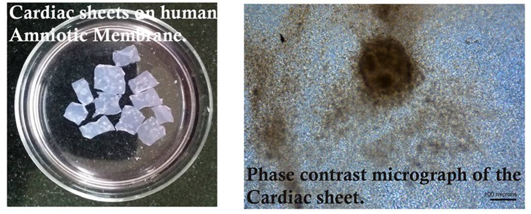
A research team from India has successfully grown human-induced pluripotent stem cell (hiPSC)-derived cardiac muscle cells on human amniotic membrane (hAM). They found that hAM outperformed the commercial gelatine-based 3D matrix Matrigel in promoting the differentiation, function and spatial organization of these cardiomyocytes. These advances could play a role in cardiac research and represent a proof-of-concept for future preclinical studies (In Vitro Cell. Dev. Biol. – Animal 10.1007/s11626-019-00321-y).
Necrosis of cardiac tissue can occur following acute events such as heart attacks. This can result in heart failure, which represents a major cause of death worldwide. In recent years, researchers have developed several approaches for regenerating injured cardiac tissue: examples include the delivery of cardiac progenitor/stem cells, synthetic materials or a combination of both to the site of injury. In addition, a growing number of studies have evaluated the functionality of decellularized biological matrices for this purpose. In this work, Shagufta Parveen and colleagues investigated the capability of hAM to support hiPSC growth and differentiation to cardiomyocytes.
The use of biological matrices such as hAM as scaffolds represents one of the current frontiers in cardiac tissue engineering. However, complete recellularization of the substrate is often challenging. Another limitation is the lack of control of cardiomyocyte phenotype, while the generation of engraftable, fully differentiated and mature cardiomyocyte sheets still remains a challenge. Moreover, the use of synthetic materials and/or allogeneic cells is associated with graft rejection. Interestingly, human amniotic membrane has been shown to modulate the inflammation process, while the use of individual-specific hiPSCs reportedly bypasses the issue of graft rejection.
The researchers examined, for the first time, the use of hAM to support the growth of hiPSC-derived cardiomyocytes. They obtained hiPSCs by altering the gene expression of stem cells derived from human placenta. These hiPSCs were then seeded on hAM or Matrigel and subjected to biochemical stimulation by growth factors to trigger the cardiac differentiation pathway.
The team then compared the morphological and biochemical characteristics between the two conditions, as well as the expression of genes and proteins related to the different stages of differentiation to cardiomyocytes.
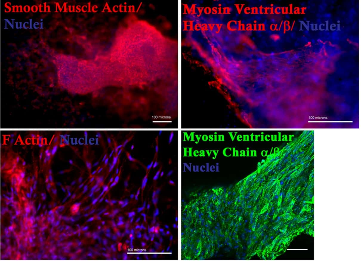
Importantly, cells grown in the presence of hAM demonstrated a higher functionality in terms of intracellular calcium transient frequency, mitochondrial organization and cellular alignmentmore closely resembling the features of cardiac muscle cells, than cells grown on Matrigel. Cells grown on hAM also exhibited an increase in the expression of protein markers related to cardiac differentiation compared with those grown on Matrigel.
The authors envision potential applications for their system within cardiac research and possibly, following appropriate preclinical studies, in clinical procedures for the treatment of heart failure.