A team headed up at Stanford University is using artificial intelligence (AI) to reduce the dose of gadolinium contrast required for MRI exams, while maintaining diagnostic quality. Recent studies have found that trace amounts of gadolinium, a heavy metal used in MRI contrast, can remain in the body after scans. The effects of this deposition are not known; thus the radiology community is working to optimize patient safety while preserving the vital information that gadolinium-enhanced MRI scans provide.
“There is concrete evidence that gadolinium deposits in the brain and body,” explains lead author Enhao Gong, who presented the study today at the RSNA annual meeting. “While the implications of this are unclear, mitigating potential patient risks while maximizing the clinical value of the MRI exams is imperative.”
To achieve this goal, Gong and colleagues are employing deep learning – an advanced AI technique. To train the deep learning algorithm, the researchers used MR images from 200 patients who had received various contrast-enhanced MRI exams. They collected three image sets for each patient: pre-contrast (zero-dose) scans; low-dose scans using 10% of the standard gadolinium dose; and full-dose scans, acquired after 100% dose administration.
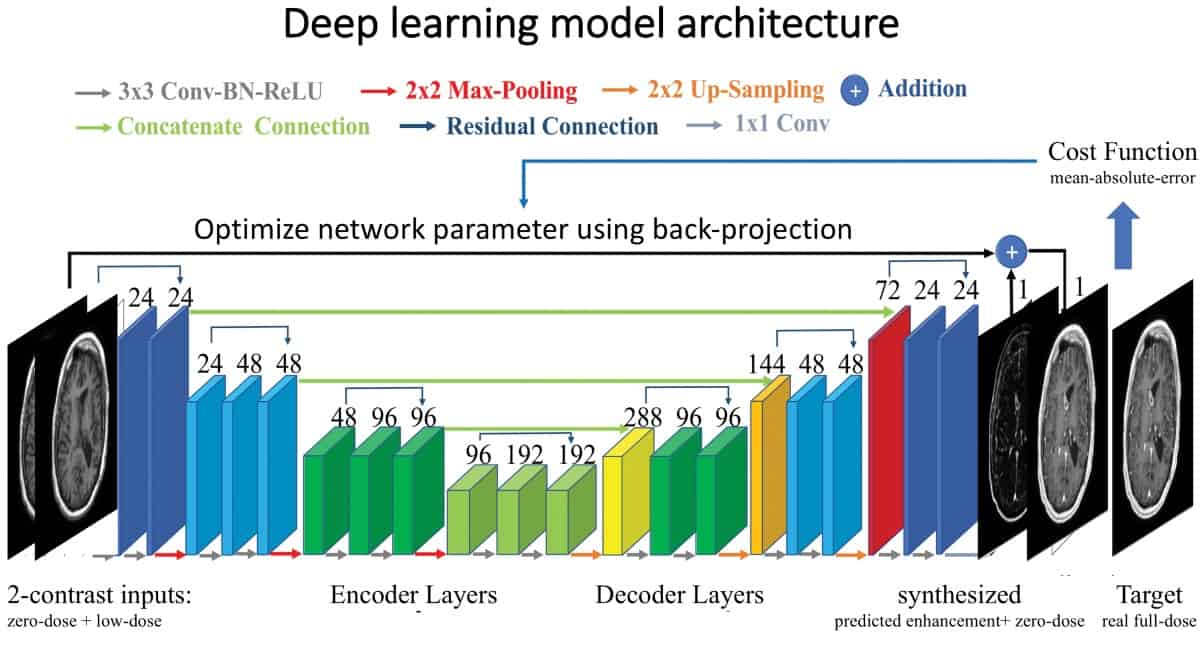
The algorithm learned to approximate the full-dose scans from the zero-dose and low-dose images. Evaluation by neuroradiologists showed that the algorithm improved image quality, with no significant different between low-dose, algorithm-enhanced MR images and full-dose, contrast-enhanced MR images. The initial results also demonstrated the potential for synthesizing the equivalent of full-dose, contrast-enhanced MR images without using any contrast agent.
The findings suggest the potential for dramatically reducing gadolinium dose without sacrificing diagnostic quality. “Low-dose gadolinium images yield significant untapped clinically useful information that is accessible now by using deep learning and AI,” says Gong.
Next, the team plans to study the method further in a clinical setting. Future research will include evaluation of the algorithm across a broader range of MRI scanners and with different types of contrast agents. “We’re not trying to replace existing imaging technology,” Gong explains. “We’re trying to improve it and generate more value from the existing information while looking out for the safety of our patients.”