The “Best-in-Physics” electronic poster session at the AAPM Annual Meeting brings together the 15 studies that scored highest in the abstract review process. These studies – categorized this year as imaging, multi-disciplinary or therapy – are considered to reflect the highest scientific quality and innovation. As always, the session proved a popular attraction and drew in large crowds of delegates keen to find out more. Here is our selection from this year’s presentations.
Imaging the scatter could track tumour motion
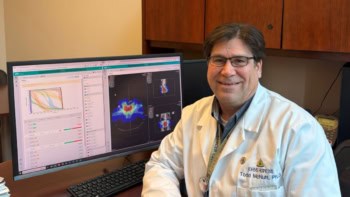
Image guidance plays a central role in radiotherapy. With this in mind, Kevin Jones from Rush University Medical Center plans to use the photons scattered out of the megavoltage beam during treatment to visualize the irradiated anatomy. “Our hypothesis is that by collecting scattered photons with a pinhole collimator, we can identify the tumour position in real time during radiation delivery,” he explained.

Jones and colleagues are developing a scatter-imaging system for tracking lung tumour motion during stereotactic body radiation therapy. He notes that one big benefit of this approach is that it doesn’t expose the patient to any additional radiation dose. It is also possible to place detectors at various positions around the patient to create a 3D image.
The prototype system comprises an off-the-shelf flat-panel X-ray detector and a pinhole collimator. The researchers optimized the collimator thickness using phantoms, and found that increasing the thickness from 7.6 to 35 mm increased the contrast-to-noise ratio by more than three times.
The team has now recorded the first scatter images collected from patients during lung SBRT. They placed the camera outside of the gantry rotation plane so it was not irradiated by the beam. Images recorded at two camera positions showed the location of the tumour. Comparison of the experimental and simulated scatter images revealed good agreement between the two at each camera position.
Jones noted that the detector – a 700 µm caesium iodide scintillator – did not collect as many photons as could a thicker detector. “The images are quite blurry, but we are optimistic that we can improve this is in the future by improving the detector and the collimator, and by moving the camera closer to the patient,” he told Physics World.
Ultimately, Jones hopes that the system will be used to track moving tumours, enabling the application of smaller treatment margins or verification of targeting accuracy. “We aim to develop an image-guided radiotherapy monitoring technique that does not require additional dose,” he said.
PEM plus ultrasound enhances biopsy targeting
Image-guided biopsy is a vital stage in diagnosis and treatment planning of breast cancers, but conventional imaging modalities have limitations. Mammography, for example, cannot easily identify lesions in dense breast tissue. Claire Park from Western University Ontario hopes to address this shortfall by using positron emission mammography (PEM) to perform high-resolution functional breast imaging.
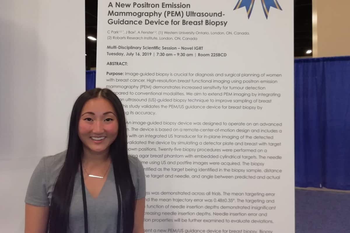
PEM offers increased sensitivity and diagnostic accuracy for tumour detection, but as it is a functional imaging approach, it doesn’t provide an anatomical reference and biopsy workflow is not yet available. “So we are combining this functional imaging with a validated ultrasound-guided biopsy technique to improve needle guidance and tumour sampling,” she explained.
Park and colleagues are developing an ultrasound-guided biopsy device that will operate with an advanced PEM system. The idea is to align the ultrasound transducer between the PEM detector plates to enable real-time needle guidance. In phantom validation studies, the biopsy device demonstrated a mean targeting error of 0.35±0.20 mm and a mean trajectory error of 0.48±0.35°. “Under ideal conditions, it can target lesions that are less than 1 mm,” she told Physics World.
The next step will be to validate the complete robotic arm, including a 3D needle tracking system. The team also plans further software developments to visualize the PEM lesion locations and determine how to accurately guide the needle to those points.
Spectral CT sees multiple contrasts simultaneously
Spectral CT using multiple energy bins enables discrimination between different high-Z materials, such as the contrast agents used in medical imaging. By employing an advanced high-flux photon counting detector and selecting energy bins above and below the K-edges of each material, several contrasts can be imaged once, as well as structures such as bone – a feat that could not be achieved using conventional CT imaging.
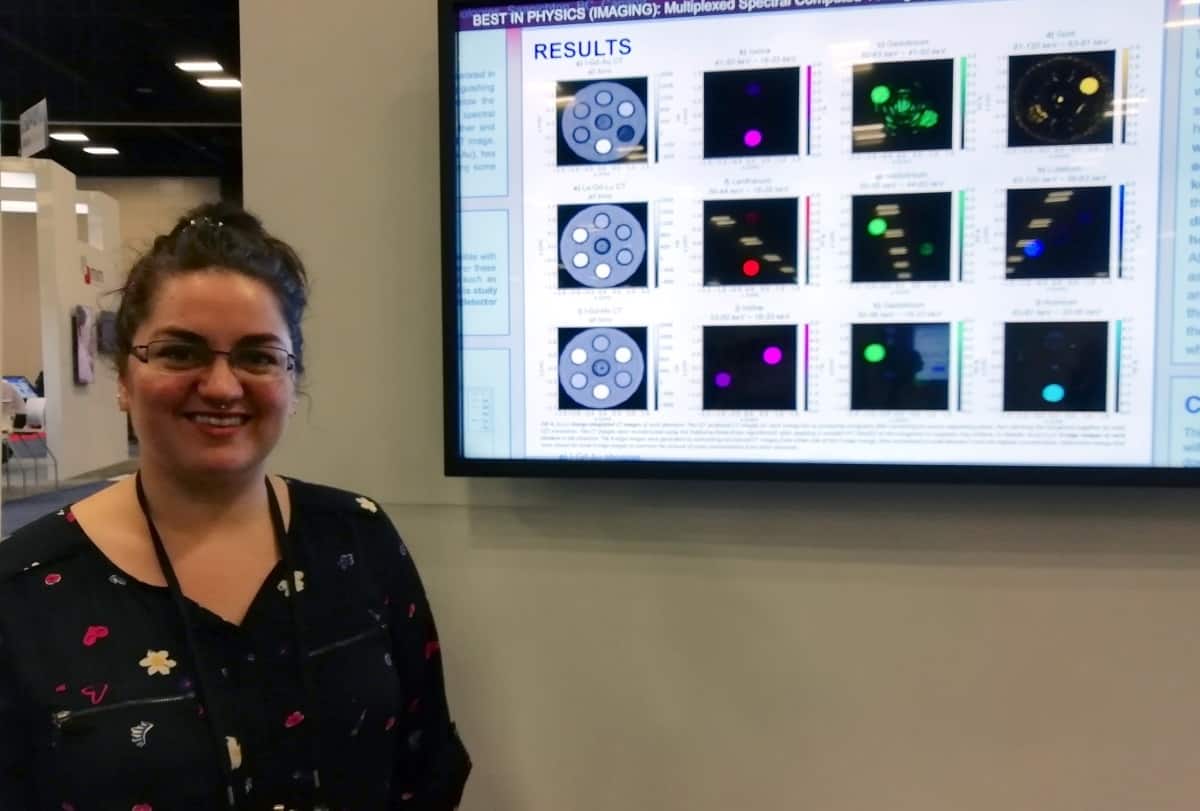
Chelsea Dunning from the University of Victoria described the use of K-edge subtraction on a spectral CT system to simultaneously image a range of contrast agents in a phantom. In addition to commonly used contrasts such as iodine (I), gadolinium (Gd) and gold (Au), she decided to also examine novel elements with emerging clinical potential. These included lanthanum (La), holmium (Ho) and lutetium (Lu), which are close in K-edge energy and may be particularly difficult to distinguish.
Dunning and colleagues 3D printed phantoms containing seven vials, one filled with water plus two concentrations of each of: Gd, I and Au; Gd, La and Lu; and Gd, Ho and I. They used a spectral CT system with a cadmium zinc telluride detector to image each phantom, choosing detector energy bins to match the K-edge energies of each component.
K-edge subtraction images of each contrast agent clearly visualized all of the different materials with minimal artefacts. The detector could distinguish three contrast agents in the La-Gd-Lu phantom, each with a difference of seven in Z and close K-edge energies.
“Most remarkably, in the phantom with iodine, gadolinium and holmium, it can discriminate all of them really well; gadolinium and holmium only differ in atomic number by three,” Dunning explained. She noted that [to their knowledge], this is the first time that contrast agents with similar Z have been imaged simultaneously.
Auto-segmentation eases radiosurgery planning
Segmentation of brain metastases for stereotactic radiosurgery planning is a time-consuming process. A team at UT Southwestern Medical Centre aims to change that. “We are developing a platform for users to upload MR images and software that performs automatic segmentation,” explained Zi Yang.

The platform imports high-resolution T1 contrast-enhanced images, and then uses a deep-learning based algorithm to carry out segmentation and labelling. At this stage, the auto-segmented brain metastases contours can be reviewed and modified by the user. The finalized contour sets are exported for use in treatment planning.
The researchers evaluated the algorithm with clinical data and found that it could perform segmentation and labelling in just 4–5 minutes, offering substantial clinical time savings and workflow improvement. Yang explained that the platform exhibits high accuracy for detection and contouring of lesions, although it currently detects some false positives. “We plan to improve the algorithm and add more functions,” she said. “Then we plan to implement the platform in the clinic.”