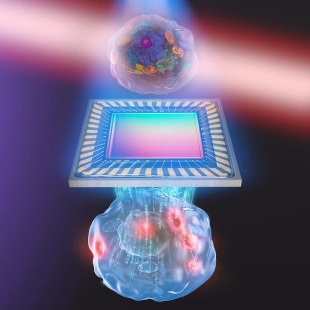
Optical imaging is widely used to image biological cells thanks to its non-destructive, label-free nature. A team of researchers in Japan has now combined two common optical techniques – quantitative phase microscopy and molecular vibrational imaging – to produce detailed images of live biological cells. The new approach could be used to observe how fundamental types of molecules are distributed in single cells, with potential applications in biology and medicine.
Quantitative phase imaging (QPI) is a technique that measures the 3D distribution of the refractive index in a transparent sample by calculating how light waves shift as they pass through the sample. It can thus be used to visualize the black-and-white outline of a biological cell as well as the outlines of the major structures inside. This information can then be used to reconstruct a 3D image of the cell.
Molecular vibrational imaging (MVI), for its part, provides information on biomolecular bonds based on Raman scattering or mid-infrared (MIR) light absorption. When molecules are excited with MIR light from a laser, they vibrate at a certain frequency and heat up their surroundings in a process known as the photothermal effect. By applying different wavelengths of MIR light to biological samples, researchers can selectively increase the temperature of specific types of chemical bonds, and so identify biostructures such as intracellular proteins, lipids or nucleic acids.
Both techniques combined
In their experiments, a team led by Takuro Ideguchi of The University of Tokyo combined QPI and MVI into a single, streamlined process. After taking a quantitative phase microscopy image of a live cell with their MIR light source turned off, they then repeat the measurement with the source turned on. The difference between the two images reveals both the outline of the major structures inside the cell and the exact locations of the type of molecule that was excited by the MIR light.
In their work, Ideguchi and colleagues say they were impressed when they first observed the molecular vibration signature characteristics of proteins, and then when this protein-specific signal appeared in the same location as the nucleolus — a structure within the cell’s nucleus in which high amounts of proteins would be expected.

Optical imaging to monitor cancer response early in the treatment process
Although it currently takes around 50 seconds or more to capture one complete image, the Tokyo team are confident that they can speed up the process by incorporating a higher-power MIR light source and a more sensitive camera into their experimental set-up.
The approach, which is detailed in Optica, could be used to study complex and fragile biological processes involved in cellular diseases and stem cell development, and to monitor drug delivery, the researchers say. In such applications, the cells need to be observed over long periods without disturbing them.


