An international team of researchers has shown that just one hour’s use of a brain–computer interface (BCI) induced spatially specific changes in the brain’s neural connections. This finding raises the possibility of creating a therapeutic system tailored for patients suffering from brain disorders or cognitive impairment (J. Physiol. 10.1113/JP278118).
A BCI is a device that collects, analyses and translates human brain signals into commands that can be understood and processed by a computer. Over the past few decades, research in this field has increased, opening up possibilities for many clinical applications. For example, a BCI that’s able to accurately translate human intentions could help restore the independence of severely disabled individuals.
One question that researchers are now keen to answer is whether these systems could have an impact on the participant’s brain. This phenomenon, in which neuronal connections are significantly affected by using the BCI device, is called BCI-induced brain plasticity.
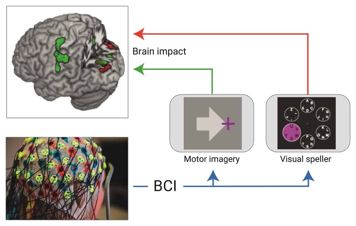
Researchers from the Max Planck Institute for Human Cognitive and Brain Sciences, TU Berlin and the Public University of Navarra have attempted to answer this exact question. In their study, they looked at whether two different types of BCI systems, one involving imagining physical movements and one involving visual stimulation, can cause signs of neural plasticity. MRI scans taken after one hour of using either of the two BCI approaches showed both structural and functional changes in the brain areas controlling these actions.
Decoding the brain
The team designed a pipeline with structural and functional brain MRI scans recorded before and after the BCI sessions. Each MRI session involved acquisition of an anatomical scan, a functional MRI (fMRI) scan with a motor imagery task and three resting-state functional MRI (rsfMRI) scans. The team chose to perform functional scanning as it reveals areas of higher brain activity, by detecting changes associated with blood flow and metabolism. The fMRI acquisitions were task-based, while the rsfMRI scans were task-free and enabled analysis of functional connectivity.
The BCI session involved the use of an electroencephalogram-based BCI system that measures and translates brain signals into commands that can be understood by a computer program. One group of 21 volunteers underwent a motor-imagery experiment, while the other group, which consisted of 19 volunteers, had to perform a visual-spelling task. During the motor-imagery session, the subjects were instructed to imagine moving their right hand or feet, while during the visual-spelling sessions, they had to spell out phrases through visually picking out letters shown on a computer screen.
Clear differences of brain activity
After collecting all the data, the team analysed the MRI scans to see whether the BCI sessions had affected the brain’s structure and function. The results were promising: in the visual-spelling group, structural MRI scans showed signal increases in the grey matter of occipital and parietal areas (brain regions involved with visual tasks); in the motor-imagery group, the sensorimotor cortex showed significant differences from the control scan.
The researchers also analysed the rsfMRI data for post-BCI session variations and observed significant clusters of modulated functional connectivity in the respective cortical areas. Finally, they note that the task-based fMRI scans recorded for both groups showed no significant differences amongst the visual-spelling volunteers, but for the motor-imagery group, showed an increase in brain activity in the left sensorimotor cortex in scans where the subjects were asked to imagine moving their right hand.
The results show that an hour of BCI training, on subjects who have never been exposed to this technology, has an effect on the brain’s structural and functional plasticity. More work is needed to figure out whether these changes translate into long-term consolidation, but for now, the emerging question is whether these devices can be used for subject-specific therapy.
As this study revealed the high spatial specificity of the BCI effects, the authors think that there is potential in “tailoring BCI-based therapeutic approaches individually to, for example, stroke patients, according to the individual patient’s lesion location”.


