A new, real-time technique to measure the changes in chirality of biomolecules in the deep ultraviolet part of the electromagnetic spectrum could be important for improved drug development and applications in medicine. The method, which is based on time-resolved circular dichroism spectroscopy, could also further our understanding on how structural changes affect the function of biomolecules.
Chiral molecules come in two versions called enantiomers, which are mirror images of each other – much like a pair of human hands. Chirality is a property related to the 3D structure of a molecule and it is crucial for the biochemical function of biomolecules. Indeed, biological processes are “homochiral”, which means that they are highly selective for when it comes to the handedness of the molecules involved. For example, most amino acids found in living organisms are left-handed, whereas most natural sugars are right-handed.
Chirality is also an important property of drugs and enantiomers of the same molecule can have entirely different chemical and biological properties. Distinguishing between them is thus particularly of interest to those developing new pharmaceuticals.
Circular dichroism spectroscopy
The main way to detect different enantiomers is using a technique called circular dichroism (CD) spectroscopy, which makes use of the fact that left-handed and right-handed molecules absorb circularly polarized light differently. CD spectroscopy is most commonly used at wavelengths below 300 nm, where biomolecules such as amino acids, DNA and peptide helices absorb most light. However this technique is challenging to perform on the time-scales of less than one picosecond at which molecules undergo structural changes that affect their chiral properties.
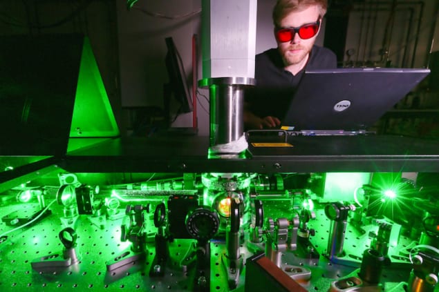
A team of researchers led by Majed Chergui of the Lausanne Centre for Ultrafast Science at the Ecole Polytéchnique Fédérale de Lausanne (EPFL) has now developed a new way to observe the chiral activity of biomolecules using a modified CD spectrometer that works in the deep-UV range (250-370 nm) with a resolution of 0.5 picoseconds.
Light waves are oscillating electromagnetic fields and when they are circularly polarized – that is, transformed to oscillate like a circular spiral around the direction of their propagation – they can sense molecular chirality, explains study lead author Malte Oppermann. A chiral molecule will thus always absorb one of the spiral directions (left- or right-handed) more than the other.
Ultrashort laser pulses
“The innovation in our instrument is that instead of using continuously radiating lamps – like in commercially available CD spectrometers – we adapted the technique so that it can use ultrashort laser pulses (with durations shorter than 0.5 picoseconds). This allows the instrument to take series of extremely fast snapshots of a molecule’s chirality as if it was a video camera capable of taking two million frames per second.”
The laser pulses employed are unique in their extreme bandwidth in the deep-UV, continuously spanning wavelengths of between 250 and 370 nm, which allows us to track chirality ranges in the range where amino acids and DNA nucleobases absorb, he tells Physics World. “Technically speaking, our instrument is special since it can record a complete CD spectrum over the whole wavelength range of our laser pulses from laser shot to laser shot. In commercial instruments, a CD spectrum has to be scanned wavelength by wavelength, which is more time consuming.”
Unearthing previously inaccessible details
Static CD spectroscopy is already a popular technique in analytical biochemistry and pharmaceutical research to investigate the structure and function of many classes of molecules, he explains. These properties are dynamic, however and can change quickly depending on the molecule’s environment – through changes in temperature, pH, chemical activity or light absorption, for example. “We are now able to observe the extremely fast time scales on which these changes occur, something that we hope will lead to new insights into how molecules, like proteins, regulate their function through their structural changes. This will be important for understanding biological activity such as protein folding, for example, or how they bind to drugs.
Since there are only very few experimental techniques that can measure structural changes that occur on these very fast time scales, the researchers say that they now hope to unearth details that were not previously accessible. Indeed, they will now use their new instrument to study myoglobin and haemoglobin. The structures of these molecules change on the picosecond timescale when they bind or eject oxygen.
“We also want to measure the real-time and ultrafast motion of molecules, such as molecular motors, for example, that can be driven and controlled by flashes of light,” says Oppermann. “These motors were the subject of research that won the 2016 Nobel Prize in Chemistry.”
Full details are reported in Optica doi.org/10.1364/OPTICA.6.00005.


