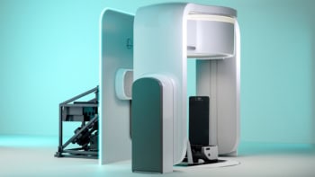
Researchers in the UK have built a prototype X-ray scanner that uses radiation at multiple wavelengths to create detailed 3D images in “colour”. The team claims that the new scanning technique could be used in a range of applications including medicine, security scanners and aerospace engineering.
Modern computed tomography (CT) scanners create high-resolution 3D images of the human body and other solid objects using a beam of X-rays with one specific wavelength. However, the technique could be improved upon by using a polychromatic beam of X-rays with a range of different wavelengths, which would allow the identification of different tissue types.
Now, researchers at the University of Manchester in the UK have developed a scanner that uses all of the wavelengths within a polychromatic X-ray beam to do just that (J. R. Soc. Interface doi: 10.1098/rsif.2007.1249).
Technology challenge
Building such a system called for the development of two key components: an array of collimating tubes to guide X-rays scattered from the sample and a pixellated, energy-sensitive detector to detect the collimated beam of scattered X-rays. Working with researchers at the University of Cambridge, UK, Robert Cernik and colleagues at Manchester used laser drilling to create the collimators. A spectroscopic-grade detector was fabricated in collaboration with the UK’s Rutherford Appleton Laboratory and Daresbury Laboratory.
The prototype device comprises a 16 × 16 array of 50 µm-diameter collimators, plus a corresponding 16 × 16 array of 300 µm2 wavelength-sensitive silicon pixel elements. To create a colour X-ray image, a polychromatic beam is incident upon the sample and the X-rays are scattered into the collimator array, which filters and directs the beams onto the detector.
As each pixel only samples a small volume, defined by the intersection of the incident and diffracted beams, the resulting images offer a high spatial resolution. The use of an array-based detector reduces the time taken to record each image to just a few minutes, lowering the radiation dose to the sample, or ultimately the patient.
The total scattering count at each pixel is used to create a 3D image of the sample. “Three-dimensional imaging is achieved by moving the sample through the fan-shaped beam in one simple z motion,” Cernik explained. “The detector and collimator assembly give you the spatial resolution in the x (the depth of the sample) and y directions.”
Meanwhile, each pixel also records the X-ray diffraction pattern as a function of wavelength from each intersection volume in the sample. This information can then be used to identify the material present at that particular point. In addition, the scattered spectra contain X-ray fluorescence information that can also be used to characterize the material.
The team tested the system on a variety of samples, starting with a thin polymer sheet marked with a test cross. When imaged using a 5 × 5 mm X-ray beam, the cross was clearly visible. The researchers also imaged a section of deer-antler bone. The system was able to image strut-like tissue structures in the bone called trabeculae and provide some information about their composition.
Cernik anticipates that one of the main medical applications of the technique will be to distinguish normal from abnormal tissues.


