Researchers in Japan are developing a medical camera that can simultaneously detect radioactive tracers used for PET and SPECT scans. The team has now demonstrated that the camera can image both tracers at the same time in a human volunteer (Phys. Med. Biol. 10.1088/1361-6560/ab33d8).
PET and SPECT scans are employed for a range of diagnostic applications. The procedure involves delivering a radioactive drug to the patient, which is then detected by the respective scanner to create an image of the patient’s internal organs. PET scans detect gamma rays with a specific energy of 511 keV, while SPECT can only detect lower-energy gamma rays because the collimators used in SPECT become transparent at higher energies.
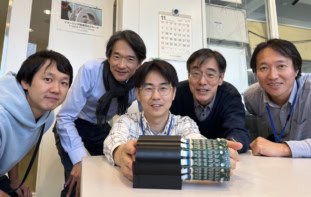
Performing separate PET and SPECT scans is time consuming and exposes the patient to increased radiation levels. As such, a team led by Takashi Nakano at Gunma University Heavy Ion Medical Center is working to combine the two procedures. The resulting system could enable doctors to scan patients in shorter times, while reducing radiation exposure.
The enabling technology is the use of a Compton camera, which can detect gamma rays in both low- and high-energy ranges without the need for collimators. This offers the potential for dual- or multi-energy radioisotope tomography, which remains challenging for conventional nuclear imaging modalities such as PET and SPECT.
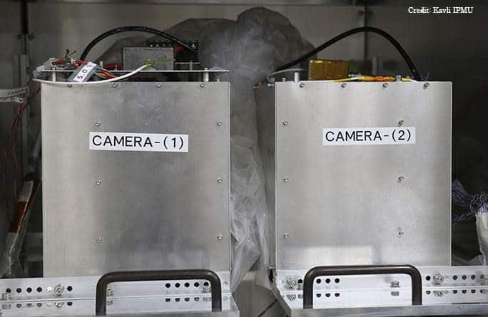
The researchers – also from Kavli IPMU, the National Institutes for Quantum and Radiological Science and Technology (QST) and the Japan Aerospace Exploration Agency (JAXA) – developed a silicon/cadmium telluride (Si/CdTe) Compton camera based on high-resolution CdTe semiconductor imaging devices. The medical camera was adapted from technology originally designed by Tadayuki Takahashi’s team at JAXA to study cosmic gamma rays.

In-human study
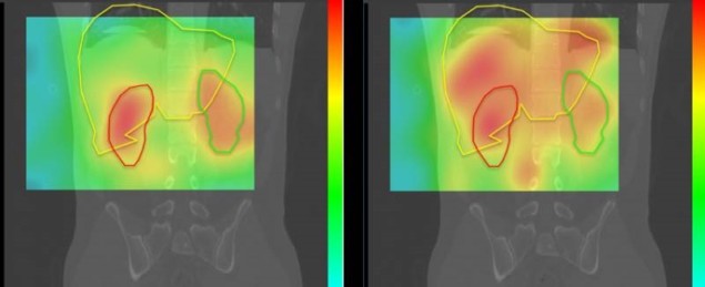
To test the Compton camera, the team imaged a volunteer using two of the most common PET and SPECT tracers: 18F-FDG and 99mTc-DMSA, respectively. On day one, they acquired CT images around the liver and kidneys. On day two, they injected the volunteer with the two tracers and recorded images from the Compton camera placed under the right side of their body.
Using a new image reconstruction algorithm to analyse the data, the researchers simultaneously created 2D images of the two radioisotopes. A map of DMSA accumulation revealed two areas of high concentration. Based on the reference CT images, these were attributed to the left and right kidneys, in agreement with the known observation that DMSA tends to accumulate in healthy kidneys. The FDG was broadly distributed over regions including both the liver and kidneys, similar to that seen in routine clinical FDG PET/CT scans.
The team points out that although the imaging resolution of the prototype Si/CdTe Compton camera is not sufficient for clinical use, the results indicate the potential of Compton cameras for future multi-energy radioisotope tomography. In addition, as the spatial resolution of a Compton camera is proportional to the detector–source distance, reducing this distance from the 300 mm used in this study would improve the resolution.
The researchers also note that the doses administered in this study (30 MBq for DMSA and 150 MBq for FDG), which were sufficient to obtain clinically useful human images in 35 min, are already below the regulatory limits. Following several more trials, they are optimistic that their imaging system will lead to new forms of medical analysis. Furthermore, it could help create completely new radioactive tracers.
“Our results indicate that the Si/CdTe Compton camera has great potential for various clinical applications in the future, and may facilitate new nuclear diagnostic procedures,” the researchers conclude. “Further improvements in its detection efficiency, spatial resolution and image reconstruction algorithms are ongoing.”


