Look at the two histopathological stains below and spot the difference.
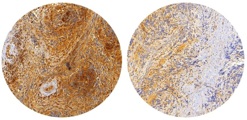
Could you tell which biopsy belongs to a high-risk neuroblastoma patient and which belongs to a low-risk neuroblastoma patient? No? Me neither. But researchers in Spain have combined top-notch image analysis with complex graph theory to identify mathematical features of these images that can help stratify tumours according to malignancy (Int. J. Cancer 10.1002/ijc.32495).
For those of you determined to guess, the high-risk neuroblastoma biopsy is on the left.
The discovery could be particularly helpful in diagnosis of the aggressiveness neuroblastoma, a type of cancer that manifests during the development of the nervous system, most commonly affecting children less than 18 months old. It is difficult to treat and patients suffer widely varying prognoses due to the involvement of many clinical and genetic factors.
The team – led by Rosa Noguera from the pathology department at the University of Valencia-INCLIVA-CIBERONC, and Luis Escudero from the University of Seville and the Seville Institute of Biomedicine-CIBERNED – first took 91 histopathological images from human neuroblastoma and isolated the distribution of a particular glycoprotein known as vitronectin (VN) in the tumour microenvironment. They had previously shown that the arrangement of VN could be connected to tumour progression (BMC Cancer 10.1186/s12885-019-5693-2).
In order to stratify neuroblastoma tumour samples according to patient risk group and the propensity for tumour cell mutation during cell division, the researchers then sought to identify topological parameters to describe the VN network. The network itself may be subdivided into territorial and interterritorial VN, with the former being located very close and within individual cells.
Topology to the rescue
The team extracted a total of 47 features with which to characterize the VN networks using an online image analysis pipeline. A mix of both morphological and topological descriptors defined each feature. The novelty of this approach comes from the inclusion of topological information – a branch of mathematics, familiar to physicists and computer scientists, that handles the preserved quantities of networks under deformation.
After analysing the data, the researchers found a statistically significant correlation between six tensegrity indices; tensegrity being one of the features they derived from the images. The researchers found that lower values of these tensegrity indices were associated with neuroblastoma tissue samples with higher risk and higher genetic instability.
The research team also note the importance of the Euler number in identifying potential neuroblastoma patients. They describe this value as “the number of objects in a sample minus the number of holes within those objects” and observed that a high Euler number per network node, in territorial VN, had statistically significant association with the high-risk patients.
Interdisciplinary teamwork
The techniques and analyses used by the research team were complicated. Fortunately, the upshot of the results is easy to understand: topological and morphological parameters associated with the VN network in histopathological neuroblastoma samples can be related to the aggressiveness of the cancer. The researchers suggest that this might be used to streamline patients prior to treatment.
Mathematical oncology: exploiting maths for cancer research
This work and its potential impact are a clear demonstration of the power of a cross-disciplinary approach to tackling cancer. By combining concepts most at home on the chalkboards of mathematics departments, the researchers have identified patterns in biology.