Cystic fibrosis is a genetic disorder in which defects in the CFTR protein (arising from mutations in the CFTR gene) can cause life-threatening symptoms in multiple organs. In the respiratory system, cystic fibrosis dehydrates the airway and produces sticky mucus in the lungs, leading to breathing problems and increasing the risk of lung infections.
One proposed treatment for cystic fibrosis is gene therapy, in which a viral vector delivers a healthy copy of the CFTR gene into airway cells to produce functional CFTR protein. To transport this vector to target cells and keep it there long enough to interact with them – key challenges for all gene therapies – researchers have coupled the vector to magnetic nanoparticles, which should allow controlled delivery to the airways using an external magnetic field.
Researchers at the University of Adelaide are now tackling another pressing challenge for successful gene therapy – visualizing the magnetic nanoparticles within live airways and manipulating them in vivo. To achieve this, they explored the use of dark-field X-ray imaging to enhance nanoparticle contrast and understand how magnetic nanoparticles move within the airway of a live rat, reporting their findings in Physics in Medicine & Biology.
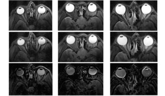
While conventional X-ray imaging relies on the absorption of X-rays, dark-field X-ray imaging detects small-angle scattering from microstructures within a sample. To perform dark-field imaging, the researchers used a 25.0 keV monochromatic beam at the SPring-8 Synchrotron in Japan. They placed a phase grid into the beam upstream of the sample, creating a pattern of beamlets at the detector. These beamlets diffuse as they scatter through the sample, and the dark-field signal can be extracted from the strength of this blurring at the detector.

“My group previously used high-resolution phase-contrast X-ray imaging for imaging nanoparticle delivery, and we were at the synchrotron when we realised the images weren’t showing the full picture,” first author Ronan Smith tells Physics World. “I developed new methods for directional dark-field imaging during my PhD, so we thought we’d see if that could help.”
Imaging nanoparticle delivery
The researchers first examined the delivery of superparamagnetic nanoparticles to an anaesthetized rat, positioned with the synchrotron beam passing through its trachea at 45°. Imaging a living animal inevitably creates background signals from the surrounding anatomy. To supress this background during nanoparticle delivery, the team employed a novel approach based on analysing the components of the directional dark-field signal.
A suspension of nanoparticles should scatter X-rays isotropically, and the major and minor scattering components of the directional dark-field signal should be equal. Asymmetric structures such as tissue, skin and hair, however, will scatter anisotropically, with most of the signal seen in the major component. By examining just the minor component, the team could enhance the contrast of the nanoparticles signal above the background.
“The directional dark-field retrieval approach was key in isolating the isotropic dark-field signal, generated by nanoparticles entering the airways, from the overlying directional dark-field signal generated by the surrounding anatomy,” Smith explains. “No one has taken this approach before as far as I know.”
Smith and colleagues delivered the nanoparticles into the rat’s trachea over 25 s, capturing 180 frames during this time, guided by the animal’s breathing. Initially, a diagonal line appeared in both the X-ray transmission and dark-field images, showing the nanoparticles starting to flow from the delivery tube into the trachea. At 22.91 s, the minor dark-field signal revealed a noticeable feature in the lower half of the tube, which became gradually clearer before being pushed out by an air bubble at the end of the delivery. The dark-field signal captured this event with 3.5 times higher signal-to-noise ratio than the transmission signal.

Imaging the delivery process revealed that the nanoparticles unexpectedly settled inside the delivery tube, with many only reaching the trachea during the last 10% of the delivery. The researchers note that this could lead to suboptimal cellular uptake of viral vectors being delivered by nanoparticles, adding that this process could not have been observed without dark-field imaging.
Rotating nanoparticle strings
Next, the team exposed the rat to a 1.17 T magnet, which caused the nanoparticles to form into string-like structures, and rotated the magnet around its trachea. With the magnet above the rat, transmission images showed that the strings were aligned vertically. As the magnet moved, the strings remained aligned to the magnetic field, suggesting that dynamic magnetic fields could indeed manipulate nanoparticles in situ.
Spectral and phase-contrast CT combine strengths to enhance X-ray imaging
With the magnet alongside the rat (partially aligning the strings along the beam axis), the strings also produced a directional dark-field signal. However, this signal was not clearly visible when the particles were aligned vertically, likely due to the beam passing through fewer nanoparticles in this position.
Smith says that the biologists in his group are now using these imaging results to enhance their work on airway gene therapy. “It’s a cyclic development process, so we have more synchrotron experiments planned to answer the questions that their results give, using a mixture of phase-contrast and directional dark-field imaging,” he explains. “We are also looking at other respiratory applications of dark-field imaging.”