Chest radiography is the most common imaging exam used for lung cancer screening. However, the size, density and location of lung lesions make their detection on chest X-rays challenging. Recently, machine-learning methods have been developed to help improve diagnostic accuracy, with deep convolutional neural networks (DCNNs), showing promise for chest radiograph interpretation.
A study from four medical centres on three continents has now demonstrated that DCNN software can improve radiologists’ detection of malignant lung cancers on chest X-rays (Radiology 10.1148/radiol.2019182465).
“The average sensitivity of radiologists was improved by 5.2% when they re-reviewed X-rays with the deep-learning software,” says Byoung Wook Choi from Yonsei University College of Medicine in Seoul, Korea. “At the same time, the number of false-positive findings per image was reduced.”
In the multicentre study, two radiologists randomly selected 800 chest X-rays from the four medical centres, including 200 normal chest scans and 600 scans with 1–3 malignant lung nodules, as confirmed by CT or pathological examination. The lung cancer scans included 704 confirmed malignant nodules (78.6% primary lung cancers and 21.4% metastases), with just over half between 1 and 2 cm in size and the remainder between 2 and 3 cm.
Next, a separate group of three radiologists from each institution interpreted the 800 X-ray scans. The group included three resident radiologists, plus four chest radiologists with five years of experience and five with more than 10 years of experience. The readers independently analysed their centres’ radiographs without clinical information, prior radiographs or CT scans.
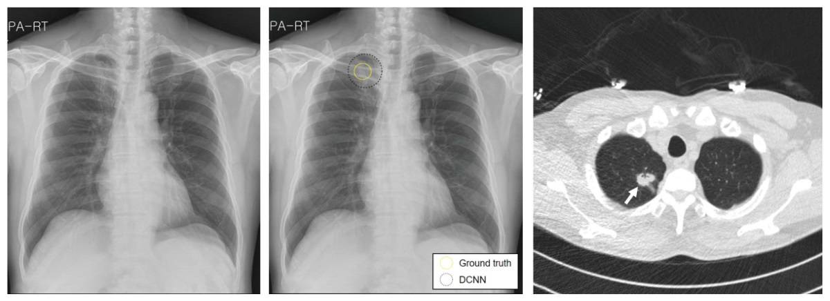
The radiologists then re-read the same X-rays with the assistance of DCNN software, which was trained to detect lung nodules. On average, the DCNN took 4.8 s to process a radiograph. When the software independently analysed the radiographs, the overall sensitivity and false-positives per image were 67.3% and 0.2, respectively.
When aided by the DCNN software, the average sensitivity of the radiologists for nodule detection improved from 65.1% to 70.3%. The number of false-positive findings per X-ray declined from 0.2 to 0.18 when the radiologists re-reviewed radiographs with the DCNN software. These trends were independent of reader experience, nodule characteristics or the vendor of the radiography system.
Hybrid machine-learning tool spots malignant lymph nodes
Use of the DCNN software resulted in a positive change (from false-negative to true-positive, or from false-positive to true-negative) in 104 of 2400 radiographs. In 54 cases, however, the decision was negatively impacted after use of the software.
“Computer-aided detection software to detect lung nodules has not been widely accepted and utilized because of high false positive rates, even though it provides relatively high sensitivity,” says Choi. “DCNN may be a solution to reduce the number of false positives.”


