A new atomic-scale spectroscopy technique has produced 3D images of a 27-atom cluster with a spatial precision of less than 0.1 nm. Created by Tim Taminiau and colleagues at QuTech of Delft University of Technology in the Netherlands and Element Six in the UK, the technique uses a defect in the crystal structure of a diamond and could be extended to determine the structures of complex individual molecules.
Nuclear magnetic resonance (NMR) spectroscopy has long been a powerful tool for the study of chemistry, biology and materials. It works by measuring the characteristic signals produced by the nuclear spins of target atoms in samples that are subjected to strong magnetic fields and resonant radio waves. As well as revealing the presence of target atoms, changes in NMR signals caused by surrounding atoms provides important structural information about a sample.
Normally, the technique involves averaging the signals produced by macroscopic samples, but recent advances have allowed isolated nuclear spins and interacting spin pairs to be identified. However, the situation is more complicated for individual molecules and nanostructures, which can comprise large clusters of nuclei. This means that many more interactions between nuclei must be characterized, which involves the formidable challenge of making very precise measurements of complex spectra of weak NMR signals and then untangling the results.
Magnetic sensor
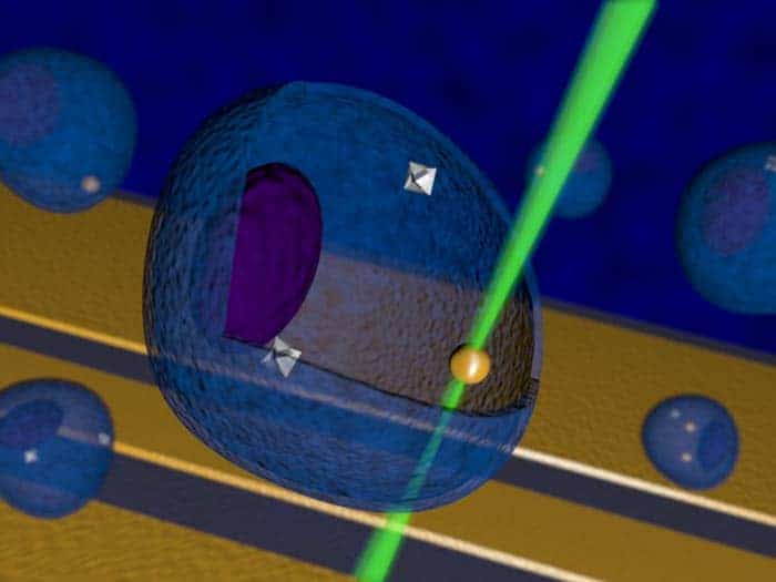
The QuTech team has addressed this challenge using a “nitrogen vacancy” (NV) centre, in which two adjacent carbon atoms in a diamond crystal are replaced by a single nitrogen atom. An NV centre is essentially an electron spin with a long coherence time and a spin state that can be measured very efficiently using laser light. This set-up can be used as an extremely sensitive magnetic sensor that can make NMR measurements on nearby nuclei.
Taminiau and colleagues developed a NV-based NMR technique for imaging a cluster of 27 carbon-13 atoms within a diamond sample. Most carbon atoms comprise carbon-12 nuclei, which have zero nuclear spin (and hence no NMR signal), whereas carbon-13 has a nuclear spin of 1/2.
The carbon-13 atoms were distributed randomly in a tiny region surrounding the NV centre. The new technique measures the couplings between pairs of nuclear spins using a three-step process. First, the NV spin is used to polarize one nuclear spin in a pair (called the probe nuclear spin). Then two radio-frequency signals are applied to couple the probe spin to the other nuclear spin in the pair – called the target. Finally, the effect of this coupling on the probe is measured using the NV spin.
Nanodiamond thermometer takes temperature of biological cells
After measuring pair-wise couplings within the cluster at high accuracy and high resolution, the team used algorithms to build up 3D images of the structure to a precision of better than 0.1 nm. This is smaller than the distance between neighbouring carbon atoms in diamond.
The next step for the Taminiau’s team is to use the technique to study nanoscale structures located outside of diamond – which the group is trying to do by creating NV centres just below the surface of diamond. If successful, the technique could soon enable researchers to gather accurate 3D images of structures including quantum devices and large, complex proteins for the first time.
The research is described in Science.