In materials science an X-ray source that is hard and bright can open the door to a wealth of detail. At Diamond Light Source X-ray beams with wavelengths of 0.1–0.2 nm outshine the Sun by a factor of 10 billion. As the 7000th paper reporting results from experiments at Diamond Light Source went to press, Physics World took a look at what the facility offers for materials scientists working on samples from ice-cream and Rembrandt fragments to next-generation data-storage technologies, as well as some of the recently opened complementary instruments for the biological and physical sciences communities on site.

Tools that image tiny structures can come in huge packages. The toroidal-shaped building housing the Diamond Light Source synchrotron has a circumference of 783 m, yet the samples it probes measure less than millimetres, and its imaging resolution reaches atomic-level detail. The facility opened in 2007 and is the largest medium-energy synchrotron in the world. It offers 31 beamlines tailored for imaging at extreme temperatures and pressures; probing electronic and magnetic materials at the atomic level; resolving the structure of complex biological samples; mapping the chemical composition of complex materials with microfocus spectroscopy; and nanoscale imaging.
The name is linked to the characteristics of the X-rays produced – hard and bright like a diamond. Here hard means in the higher-energy part of the X-ray region of the electromagnetic spectrum, so smaller structures can be resolved. To produce the X-rays, electrons produced through thermionic emission from a cathode are accelerated in a linear accelerator and then a booster synchrotron where they reach close to the speed of light, before circulating in a storage ring around 560m in circumference. Magnets direct the electron beam around the storage ring, and each time the electron beam passes through these magnets it emits synchrotron radiation. The parameters of this radiation, such as the spectral wavelength and polarization, are tightly controlled to optimize for different experiments, and high-specification sample mounting and metrology equipment also contribute to high-resolution data mapping.
Saving Homer

Stephen Price, a researcher at Finden and formerly a Diamond Light Source scientist, is currently working with the Rijksmuseum in Amsterdam on a sample of Rembrandt’s painting “Homer”, which dates back to 1663. As he explains, the sample is of a white bloom or crust that forms on the painting despite the best efforts of conservationists. His aim has been to identify the chemistry of the crust and hopefully determine how to prevent such crusts forming.
“Normally a lab-based diffractometer has quite a large beam profile of a few millimetres in size and the sample here is much less than one millimetre,” says Price, explaining that even if a lab-based X-ray source could identify the phases there would be no spatial information as to whether those phases were at the surface, middle or right by the canvas. “Further to that a lab-based source just doesn’t have the flux to image such a small volume,” he adds.
Using the microfocus X-ray beam at Diamond to scan the sample at different angles, they were able to show how the lead paint had reacted with atmospheric pollutants including sulphur dioxide, which was forming the white crust disfiguring the painting. “Using this information the conservation team at the Rijks can investigate further how to prevent and reverse this degradation process,” says Price.
Shedding new light on old art
Read more

Shedding new light on old art
The perfect slurp
As well as high-end art Diamond Light Source has made contributions to more gastronomic aspects of culture, namely what makes ice-cream so good. Ice-cream typifies food where the feel and texture contribute as much – if not more – to the experience as the flavour itself. As a result the perfect ice-cream hinges on microstructural characteristics deeply embedded in materials science.
Above -30 °C the subtle balance of crystal and bubble size versus the unfrozen matrix starts to disintegrate, hence the disappointment of eating ice-cream where the storage temperature has cycled between hot and cold in a process described with no exaggeration as “thermal abuse”. X-ray studies of ice-cream that had been thermally abused in 14 cycles could show that the ice crystals and air bubbles grew, and that they grew by less after 7 cycles. However, to understand the interaction between different microstructural features required in situ studies during the cycling itself.
“This work also revealed other interesting phenomena, including the role of the unfrozen matrix in maintaining the ice cream’s microstructural stability and the complex interactions between ice crystals and air bubbles,” explains Peter Lee, Acting Director at the Research Complex at Harwell (RCaH) next to Diamond, and one of the researchers who played a leading role in these results. “For example, the melting and recrystallization of ice crystals significantly affect the air bubbles’ morphology and the behaviour of the unfrozen matrix.”
A twist in skyrmion data storage prospects
Magnetic materials have been widely used for computer storage for decades but as devices shrink and memory requirements intensify, there has been keen interest in possible alternatives, a recent candidate being magnetic skyrmions. In these nanoscale magnetic quasiparticles, the field vectors follow twisted vortices pointing towards or away from a single point in space. Being small, stable and responsive to their environment, they have attracted a lot of interest for next-generation data storage technology as well as spintronics, but questions remain as to how surfaces affect skyrmions.
Using resonant elastic scattering of circularly polarized X-rays at Diamond, Shilei Zhang at the University of Oxford, alongside colleagues in the UK, Germany and Switzerland were able to retrieve information about changes in the type of twisting in the skyrmions over several hundred nanometre distances from the surface. “This has far-reaching implications for the creation of skyrmions in surface-dominated systems and identifies, more generally, surface-induced gradual variations deep within a bulk material and their impact on tailored functionalities as an unchartered scientific territory,” says Zhang.
The results were published in the Proceedings of the National Academy of Science in the 7000th paper reporting results from experiments at Diamond Light Source. “Diamond’s 7000th publication exemplifies the links between fundamental research, applied science and the technologies that move humanity forwards,” says Laurent Chapon, Diamond’s Physical Sciences Director.
Precision support technology
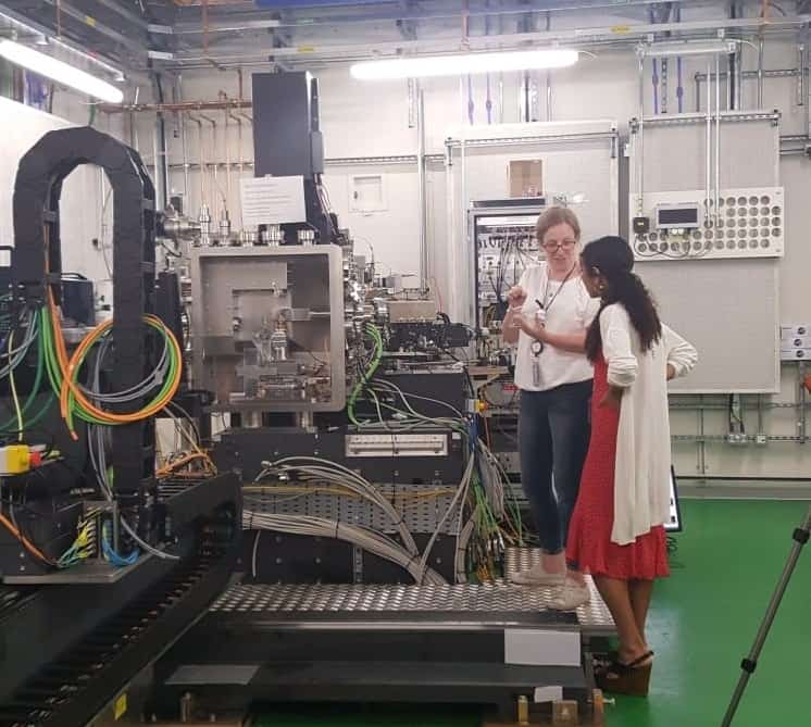
The beamline’s optics hutch, where the light is focused and filtered, can be situated a substantial distance away from the experimental hutch where the experiments take place. Julia Parker, who works on the beamline for nanoscale studies mounts samples in a hutch that is 185 metres away from the hard nanoprobe beamline’s optics hutch to maximize the distance from the focusing optic to the sample. The beams travel to the hutch along vacuum lines with negligible attenuation, and users can control all the parameters from the control cabin next to the experimental hutch. “You can even change parameters from home if you wanted to,” adds Parker.
The hard nanoprobe beamline has a beam size of 50 nm and opened for users in March 2017. It allows users to obtain chemical and structural information with 50 nm resolution, using beams with energies ranging from 5-23 keV, and accommodates in situ measurements under strained, wet and heated conditions for both organic and inorganic samples.
While the technical specifications of the facilities at Diamond are impressive, improvements are ongoing. One recent development was the introduction of a device that can step through angles one nanoradian at a time – equivalent to the angle a telescope on Earth would move through to look at the toe or the heel of a footprint on the moon. The device enables better testing and inspection of the mirrors that focus the X-ray beams and exemplifies the level of accuracy of the equipment at Diamond that enables such high-precision experiments.
State-of-the-art electron microscopy
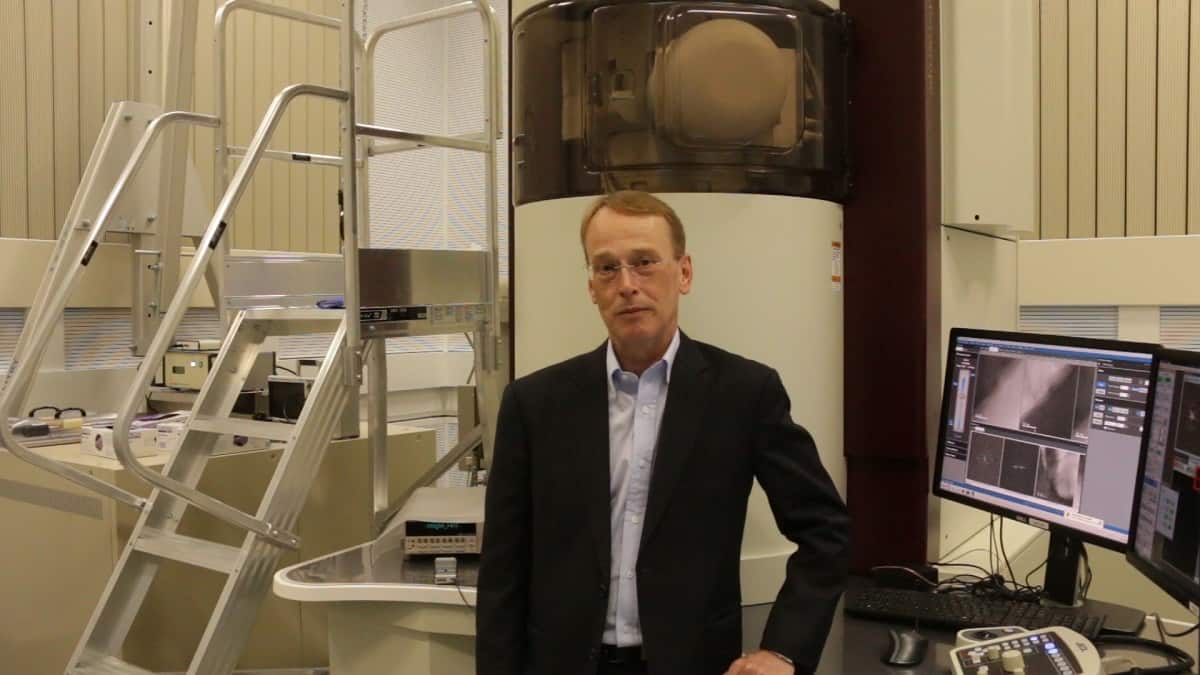
In 2016 Diamond announced the opening of an electron microscopy imaging centre specifically geared towards the needs of the physical sciences community. The electron Physical Sciences Imaging Centre (ePSIC) is a collaboration between Diamond, the University of Oxford and the catalyst company Johnson Matthey plc, and is available to users free of charge on a peer-reviewed proposal basis. It is home to two state-of-the art transmission electron microscopes one running from 200 kV and the other from 300 kV and capable of resolving features just 47 picometres in size. As Parker points out these complementary facilities are very useful for the nano beamline experiments where the size of samples is one of the main challenges. The facilities also attract researchers running projects that do not use the main X-ray source.
“We provide a very high-performance instrument and also expert staffing so that users who don’t have the expertise to drive the instrument themselves can come here and our staff scientists will run the experiment for them,” explains Angus Kirkland, Professor of Materials at the University of Oxford and Science Director at the Centre.
Scanning transmission electron microscopy explained

Studies for the nuclear industry
Experiments at ePSIC include studies of 2D materials, and materials for catalysts, batteries and other energy applications, as well as steels for aerospace and nuclear industry materials. Chris Grovenor, a Professor of Materials at the University of Oxford, uses the electron microscopes at ePSIC to study corrosion in zirconium alloys, which are the preferred cladding alloy in the nuclear industry for pressurized water reactors. Their role is to separate the water from the uranium dioxide fuel, as any mixing between the two can be incredibly dangerous. Inside the reactor the zirconium is subject to intense heat and pressure as well as neutron damage, which causes it to corrode with the formation of an oxide layer.
At present the industry acts in line with very conservative estimates as to how long the rods can be left in the reactor before corrosion embrittles the cladding, so only between 30% and 40% of the uranium is burnt. “Every time you switch the reactor off for a day it costs a million pounds on account of the energy it is not producing,” explains Grovenor, adding that sufficient knowledge to safely leave the rods in for longer would also reduce nuclear waste. He and his research team are studying how the oxide layer forms at the atomic scale to better understand how long the zirconium can survive the reactor conditions before it needs replacing.
Developments at ePSIC
After ten years or so of rapid development the hardware of the microscopes has plateaued. Now as Staff Scientist Chris Allen points out the interesting bit is what to do with the hardware, and here increasingly the focus is on in situ experiments, where imaging takes place in temperature and pressure conditions closer to those in which a catalyst operates, for example.
There is also a lot of interest in high-speed imaging – 4D TEM – to delve into fast dynamical processes, such as the evolution of defects and dopants. Thanks to one of Diamond’s spin-off companies, which have developed ultrafast imaging cameras, the scientists at ePSIC have had access to the technology very early on. “There’s not many labs internationally that have access to these cameras,” says Allen. As camera frame rates increase, the simulation timescales that computational chemists can reach are also increasing and are approaching milliseconds. “So we are potentially getting to the stage where we can directly compare what the computational chemists are saying about the life times of various features with our imaging, and that for me is really exciting.”
Biological samples out in the cryogenically cold
So where do these world-class facilities for imaging alloys and 2D materials leave scientists working on biological samples? Catering specifically for this community, in June 2015 Diamond brought online its first microscope as part of the Electron Bio-Imaging Centre (eBIC) following the award of a £15.6 million grant from the Wellcome Trust, the Medical Research Council (MRC) and the Biotechnology and Biological Sciences Research Council (BBSRC). The centre is now home to several world-class microscopes, including cryo-electron microscopes for imaging cells and other frozen hydrated biological samples, and staff scientists can assist users in both single particle analysis and cryo-tomography of their samples.
Having the two electron microscope centres on site at Diamond provides a full suite of complementary instrumentation to help researchers push the boundaries of understanding in materials science, a field that is rife with interdisciplinary developments. In the words of Diamond Science Director Laurent Chapon, “Discovery may be an everyday occurrence here at Diamond, but it never loses its shine.”
Diamond Light Source – where materials surrender their secrets